. 2010 Jan 22;6(1):e1000830.
doi: 10.1371/journal.pgen.1000830.

Differential localization and independent acquisition of the H3K9me2 and H3K9me3 chromatin modifications in the Caenorhabditis elegans adult germ line

Affiliations

Differential localization and independent acquisition of the H3K9me2 and H3K9me3 chromatin modifications in the Caenorhabditis elegans adult germ line

Jessica B Bessler et al. PLoS Genet. .

Abstract

Histone methylation is a prominent feature of eukaryotic chromatin that modulates multiple aspects of chromosome function. Methyl modification can occur on several different amino acid residues and in distinct mono-, di-, and tri-methyl states. However, the interplay among these distinct modification states is not well understood. Here we investigate the relationships between dimethyl and trimethyl modifications on lysine 9 of histone H3 (H3K9me2 and H3K9me3) in the adult Caenorhabditis elegans germ line. Simultaneous immunofluorescence reveals very different temporal/spatial localization patterns for H3K9me2 and H3K9me3. While H3K9me2 is enriched on unpaired sex chromosomes and undergoes dynamic changes as germ cells progress through meiotic prophase, we demonstrate here that H3K9me3 is not enriched on unpaired sex chromosomes and localizes to all chromosomes in all germ cells in adult hermaphrodites and until the primary spermatocyte stage in males. Moreover, high-copy transgene arrays carrying somatic-cell specific promoters are highly enriched for H3K9me3 (but not H3K9me2) and correlate with DAPI-faint chromatin domains. We further demonstrate that the H3K9me2 and H3K9me3 marks are acquired independently. MET-2, a member of the SETDB histone methyltransferase (HMTase) family, is required for all detectable germline H3K9me2 but is dispensable for H3K9me3 in adult germ cells. Conversely, we show that the HMTase MES-2, an E(z) homolog responsible for H3K27 methylation in adult germ cells, is required for much of the germline H3K9me3 but is dispensable for H3K9me2. Phenotypic analysis of met-2 mutants indicates that MET-2 is nonessential for fertility but inhibits ectopic germ cell proliferation and contributes to the fidelity of chromosome inheritance. Our demonstration of the differential localization and independent acquisition of H3K9me2 and H3K9me3 implies that the trimethyl modification of H3K9 is not built upon the dimethyl modification in this context. Further, these and other data support a model in which these two modifications function independently in adult C. elegans germ cells.


Conflict of interest statement

The authors have declared that no competing interests exist.

Figures
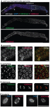
Figure 1
Figure 1. H3K9me2 and H3K9me3 localization in the adult hermaphrodite germ line.
(A) A germ line dissected from a wild type C. elegans hermaphrodite. The premeiotic region of the germ line is on the left, while germ cells at the diplotene stage of meiotic prophase are on the right. The top panel shows anti-H3K9me2 (green) and anti-H3K9me3 (red) staining overlaid on DAPI-stained chromosomes (blue). The middle panel shows anti-H3K9me2 staining alone and the bottom panel shows anti-H3K9me3 staining alone. Boxed regions are enlarged in sections (B,C). Scale bar = 10 µm. (B) Enlargement of premeiotic nuclei from A, showing that both H3K9me2 and H3K9me3 are broadly distributed, but that their peaks of intensity often do not overlap. Scale bar = 5 µm. (C) Enlargement of pachytene nuclei from A, showing that H3K9me2 is concentrated on a subset of chromosome regions, whereas H3K9me3 is more broadly distributed throughout the chromatin. Scale bar = 5 µm. (D) Examples of wild type diakinesis nuclei, in which H3K9me3 is strongly present on all chromosomes but H3K9me2 is barely detectable. Scale bar = 5 µm. (E) Examples of diakinesis bivalents stained with DAPI and H3K9me3, showing that H3K9me3 exhibits a distinct banding pattern. Scale bar = 5 µm.
Figure 2
Figure 2. H3K9me2 and H3K9me3 localization in pachytene nuclei from XO males and him-8 XX hermaphrodites.
(A) Male pachytene germ cell nuclei each have a single X chromosome. H3K9me2 is highly enriched on this unpaired X chromosome, while H3K9me3 is present on all chromosomes and is not enriched on the X chromosome. (B) him-8 hermaphrodite pachytene germ cell nuclei, which carry two unpaired X chromosomes. H3K9me2 is highly enriched on both unpaired X chromosomes, while H3K9me3 is present on all chromosomes. (C) met-2(n4256) male pachytene germ cell nuclei. In the met-2 mutant nuclei, H3K9me2 is no longer present on the unpaired male X chromosome, while the distribution of H3K9me3 appears unaffected. (D) met-2(n4256); him-8 hermaphrodite pachytene germ cell nuclei. In these nuclei, H3K9me2 is no longer present on the unpaired X chromosomes. The distribution of H3K9me3 appears unaffected. All germs cells have been co-stained with anti-H3K9me2 (green), anti-H3K9me3 (red) and DAPI (blue). Scale bar = 5 µm.
Figure 3
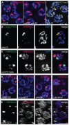
Figure 3. Arrays composed of transgenes with soma-specific promoters are enriched for H3K9me3.
(A) Left: Hermaphrodite pachytene nuclei carrying mIs10. The mIs10 array is highly enriched for H3K9me3 (red). Middle: zim-2; mIs10 hermaphrodite pachytene nuclei stained with anti-H3K9me3. mIs10 is still enriched for H3K9me3 in the zim-2 mutant despite a lack of pairing between the two copies of chromosome V (into which mIs10 is integrated). The basal level of H3K9me3 is not visible in these images due to the extreme brightness of H3K9me3-staining associated with mIs10. Right: mIs10 hermaphrodite pachytene nuclei stained for H3K27me3 (yellow). H3K27me3 is broadly distributed on all chromosomes, with a slight enrichment on the X chromosome; it is not concentrated on mIs10. Blue = DAPI. (B) mIs10 hermaphrodite pachytene nucleus hybridized with a high-copy array FISH probe (green), concurrent with anti-H3K9me3 IF (red). The brightest part of the H3K9me3 staining overlaps with the FISH signal. (C) mIs10 male germ cells co-stained for H3K9me2 (green) and H3K9me3 (red). H3K9me3 is enriched on mIs10, while H3K9me2 is enriched on the partnerless X chromosome. (D) Germ cell nuclei carrying different high-copy arrays, composed of soma-specific promoters, stained for H3K9me3 (red). All examined arrays with soma-specific promoters showed an enrichment for H3K9me3. (E) Nuclei containing the high-copy let-858::gfp transgene array ccEx7271, co-stained for H3K9me2 (green) and H3K9me3 (red). let-858::gfp is expressed in the soma but is silenced in the germ line. This array is enriched for H3K9me2, but H3K9me3 levels are indistinguishable from the surrounding chromosomes. Scale bar = 5 µm.
Figure 4
Figure 4. Distinct properties of H3K9me2 and H3K9me3-enriched domains.
(A) axIs36 male pachytene nuclei co-stained with anti-H3K9me2, anti-H3K9me3 and DAPI. H3K9me2 remains restricted to the X chromosome, while H3K9me3 is restricted to axIs36. (B) him-8; axIs36 hermaphrodite pachytene nuclei co-stained with anti-H3K9me2, anti-H3K9me3 and DAPI. Top: a nucleus where both X chromosomes carrying axIs36 stain for H3K9me2 and H3K9me3. 11 of 20 nuclei examined showed two distinct H3K9me2 signals and two distinct H3K9me3 signals. Bottom: a nucleus where one X chromosome carrying axIs36 is stained only for H3K9me3 and the other X chromosome is stained for both H3K9me2 and H3K9me3. 5 of 20 nuclei examined showed two distinct H3K9me3 signals but only one H3K9me2 signal (see text for additional information). In both (A,B), H3K9me3 staining also corresponds to the presence of a DAPI-faint region. (C) axIs36 male nucleus stained only with anti-H3K9me2 and DAPI. A gap in the H3K9me2 staining indicates the position of the axIs36 transgene array inserted into the X chromosome and also corresponds to a region of reduced DAPI intensity, demonstrating that a DAPI-faint region corresponding to axIs36 is still observed in experiments that do not include anti-H3K9me3 antibody. (D) axIs36 male nucleus co-stained with anti-H3K9me2, anti-H3K9me3 and Höechst dye #33258. These data indicate that faint chromatin staining coincident with axIs36 can be seen using Höechst as well as DAPI. (E) mIs10 hermaphrodite nucleus co-stained for anti-H3K9me2, anti-H3K9me3 and DAPI. A DAPI-faint domain corresponds to the presence of the mIs10 high-copy array. Green = H3K9me2, Red = H3K9me3, Blue = DAPI or Höechst. Scale bar = 5 µm.
Figure 5
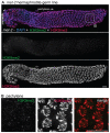
Figure 5. met-2 is required for H3K9me2 in the adult germ line.
(A) A germ line dissected from a met-2(n4256) C. elegans hermaphrodite. The premeiotic region of the germ line is on the left, while germ cells at the diplotene stage of meiotic prophase are on the right. Top: anti-H3K9me2 staining (green) and anti-H3K9me3 staining (red) overlaid on DAPI-stained chromosomes (blue). Middle: anti-H3K9me2 staining alone. Bottom: anti-H3K9me3 staining alone. In the absence of MET-2, no chromosomal H3K9me2 staining is visible. Boxed region is enlarged in (B). Scale bar = 10 µm. (B) Enlargement of pachytene nuclei from A, showing that H3K9me2 is not visible in the met-2(n4256) mutant germ line, while H3K9me3 is unaffected. Scale bar = 10 µm.
Figure 6
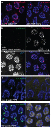
Figure 6. Effects of loss of MET-2 on high-copy arrays.
(A) met-2(n4256); ccEx9747 and ccIs4251; met-2(n4256) hermaphrodite pachytene nuclei stained for H3K9me3. H3K9me3 is unaffected in met-2 mutant worms. (B) Male met-2(n4256); him-8; axIs36 pachytene nuclei co-stained for H3K9me2 and H3K9me3. Despite the absence of H3K9me2, H3K9me3 is still restricted to the axIs36 array on the X chromosome. (C) met-2(n4256); ccEx7271 pachytene nuclei stained with H3K9me2 and examined for let-858::gfp expression. Even without MET-2 and H3K9me2, the let-858::gfp transgene is still silenced in the germ line, demonstrated here by the lack of GFP visible in germline nuclei. (D) Control hpl-2; ccEx7271 pachytene nuclei stained with H3K9me2 and examined for let-858::gfp expression. In the absence of hpl-2, H3K9me2 is still present on the array containing the let-858::gfp transgene, but let-858::gfp expression is desilenced in the germ line, as GFP is visible in the nuclei of germ cells. Red = H3K9me3, Green = H3K9me2, Yellow = GFP, Blue = DAPI. Scale bar = 5 µm.
Figure 7
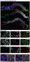
Figure 7. met-2 phenotypes in the hermaphrodite gonad.
(A) Wild type and met-2 nuclei stained with HIM-8 antibody (Green). A single HIM-8 signal in each nucleus indicates that the X chromosomes in met-2 mutant worms are paired. Blue = DAPI. Scale bar = 5 µm. (B) Wild-type and met-2 pachytene nuclei co-stained for SC components SYP-1 and HTP-3. Chromosomes in met-2 mutant worms appear to pair and synapse normally. Scale bar = 5 µm. (C) Diakinesis nuclei from met-2 mutants. The top panel represents the 99.1% of met-2 diakinesis nuclei with six DAPI-stained bodies, indicative of six pairs of homologous chromosomes held together by chiasmata. The bottom panel represents the 0.9% of met-2 mutant diakinesis nuclei with greater than six DAPI-staining bodies, indicating that one or more chromosome pairs are not linked by chiasmata. Scale bar = 5 µm. (D) Ethanol fixed wild type whole worm, showing normal organization and progression of nuclei, from the distal germ line (marked with white arrow) to the proximal end of the germ line. Scale bar = 10 µm. (E) Ethanol fixed met-2 whole worm with abnormal endomitotic nuclei (marked by white ovals). (F) Ethanol fixed met-2 whole worm with a germ line tumor (indicated by the white oval). (G) Table summarizing the quantitation of met-2(n4256) and met-2(ok2307) hermaphrodite gonad phenotypes. All images are of met-2(n4256) animals.
Figure 8
Figure 8. mes-2 is required for wild-type H3K9me3 levels.
(A) A germ line dissected from an M+Z- mes-2 C. elegans hermaphrodite. The premeiotic region of the germ line is on the left, while germ cells at the diplotene stage of meiotic prophase are on the right. Top: anti-H3K9me2 (green) and anti-H3K9me3 (red) staining overlaid on DAPI-stained chromosomes (blue). Boxed regions are enlarged in (B,C). Middle: anti-H3K9me2 staining alone. Bottom: anti-H3K9me3 staining alone. Most H3K9me3 staining is absent in the mes-2 mutant, however DAPI-stained bodies thought to be mitotic figures still stain for H3K9me3 (underlined with dashed line) and diplotene nuclei retain some anti-H3K9me3 staining (arrows). An asterisk indicates the approximate position of meiotic prophase entry, while the presence of a single line of nuclei indicates the beginning of the diplotene stage. Scale bar = 10 µm. (B) Enlargement of the premeiotic region from A, showing that H3K9me3 is still present on chromatin that appears to correspond to mitotic figures. Scale bar = 5 µm. (C) Enlarged pachytene nuclei from A, showing that while H3K9me2 is unaffected, most nuclei lack H3K9me3 staining. Scale bar = 5 µm. (D) Representative mes-2 diakinesis nuclei. Even though H3K9me3 staining is reduced or lost from the majority of germ cells, diakinesis nuclei in the mes-2 mutant usually retain H3K9me3 staining. The absence of H3K9me2 from diakinesis-stage chromosomes seen here is a normal feature of germ lines. Scale bar = 5 µm. (E) mes-2; mIs10 and mes-2; axIs36 stained with anti-H3K9me3 and DAPI. While most endogenous H3K9me3 staining is lost, the transgene arrays can retain reduced H3K9me3. Scale bar = 5 µm.

References

    1. Kouzarides T. Chromatin modifications and their function. Cell. 2007;128:693–705. - PubMed
    1. Yasuhara JC, Wakimoto BT. Molecular landscape of modified histones in Drosophila heterochromatic genes and euchromatin-heterochromatin transition zones. PLoS Genet. 2008;4:e16. doi: 10.1371/journal.pgen.0040016. - DOI - PMC - PubMed
    1. Vakoc CR, Mandat SA, Olenchock BA, Blobel GA. Histone H3 lysine 9 methylation and HP1gamma are associated with transcription elongation through mammalian chromatin. Mol Cell. 2005;19:381–391. - PubMed
    1. Kim A, Kiefer CM, Dean A. Distinctive signatures of histone methylation in transcribed coding and noncoding human beta-globin sequences. Mol Cell Biol. 2007;27:1271–1279. - PMC - PubMed
    1. Grewal SI, Jia S. Heterochromatin revisited. Nat Rev Genet. 2007;8:35–46. - PubMed

Publication types

MeSH terms