. 2010 Jan 22;6(1):e1000732.
doi: 10.1371/journal.ppat.1000732.

Placental syncytiotrophoblast constitutes a major barrier to vertical transmission of Listeria monocytogenes

Affiliations

Placental syncytiotrophoblast constitutes a major barrier to vertical transmission of Listeria monocytogenes

Jennifer R Robbins et al. PLoS Pathog. .

Abstract

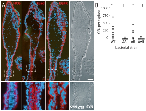
Listeria monocytogenes is an important cause of maternal-fetal infections and serves as a model organism to study these important but poorly understood events. L. monocytogenes can infect non-phagocytic cells by two means: direct invasion and cell-to-cell spread. The relative contribution of each method to placental infection is controversial, as is the anatomical site of invasion. Here, we report for the first time the use of first trimester placental organ cultures to quantitatively analyze L. monocytogenes infection of the human placenta. Contrary to previous reports, we found that the syncytiotrophoblast, which constitutes most of the placental surface and is bathed in maternal blood, was highly resistant to L. monocytogenes infection by either internalin-mediated invasion or cell-to-cell spread. Instead, extravillous cytotrophoblasts-which anchor the placenta in the decidua (uterine lining) and abundantly express E-cadherin-served as the primary portal of entry for L. monocytogenes from both extracellular and intracellular compartments. Subsequent bacterial dissemination to the villous stroma, where fetal capillaries are found, was hampered by further cellular and histological barriers. Our study suggests the placenta has evolved multiple mechanisms to resist pathogen infection, especially from maternal blood. These findings provide a novel explanation why almost all placental pathogens have intracellular life cycles: they may need maternal cells to reach the decidua and infect the placenta.


Conflict of interest statement

The authors have declared that no competing interests exist.

Figures

Figure 1
Figure 1. Comparison of in vivo placental structure to placenta explant model.
(A) Structure and orientation of fetus and placenta in uterus at ∼6 weeks of gestation. Fetal structures are represented in shades of blue and purple while maternal are in shades of red. Maternal structures: MY: myometrium, SA: spiral arteries, DD: decidua (uterine lining during pregnancy), IVS: intervillous space filled with maternal blood. Fetal structures: VT: villous tree, CP: chorionic plate, UC: umbilical cord, AF: amniotic fluid. (B) (Enlargement of boxed area in panel A) Maternal blood surrounds the villous tree composed of anchoring (AV) and floating villi (FV), which are covered by a syncytiotrophoblast (SYN) that is underlaid by subsyncytial cytotrophoblasts (sCTB) and a basement membrane. The subsyncytial CTB layer grows increasingly discontinuous in later trimesters. Gas and nutrient exchange with the maternal blood occurs across the syncytiotrophoblast to supply fetal capillaries in the stroma (STR). At the uterine wall, extravillous cytotrophoblasts (EVT) anchor the villous tree in the decidua. Some invade the decidua and move away from the tip to remodel maternal spiral arteries, with altered gene expression patterns as they move (not shown). Notably, E-cadherin expression decreases as VE-cadherin expression rises in distal (relative to fetus) extravillous cytotrophoblasts. (C) A six-week placental explant anchored in Matrigel. Bar = 1 mm. (D) Cartoon representation of the relevant structures seen in panel C.
Figure 2
Figure 2. L. monocytogenes grows variably in placental explants.
(A) Intracellular survival of L. monocytogenes in 86 explants from 18 placentas infected with ∼2×10⁶ 10403S (filled circles) or EGDe (open circles) wild type strains for 30 min. Gentamicin was added at 60min to kill extracellular bacteria and maintained in media thereafter. Infection is highly variable and growth is slower than in most cell lines. Bars = median values. (B) Number of internalized bacteria at 2 h post inoculation (p.i.) correlates with the number of anchoring villi in the explant (n = 30 explants, r2 = 0.49). (C) Histological section of explant frozen and sliced at 8 h p.i., then stained for L. monocytogenes (green), DNA (blue), and EGFR (red), which stains trophoblast membranes. Bacteria are found in extravillous cytotrophoblasts (EVT) but not syncytiotrophoblast (SYN). Matrigel (MAT) and stroma (STR) are also indicated. Bar = 100 µm.
Figure 3
Figure 3. L. monocytogenes enters the placenta primarily at invasive villus tips.
(A) Consecutive histological sections of a permissively infected explant at 8 h post-inoculation, frozen and stained for L. monocytogenes (green) and DNA (blue). Left panel and inset 1: red = cytokeratin (CK), expressed by cytotrophoblasts (CTB). In middle panel with insets 2 and 3: red = βHCG (HCG), which primarily stains syncytiotrophoblast (SYN). Subsyncytial cytotrophoblasts (sCTB) underlie the syncytiotrophoblast. Where cytotrophoblasts invade from the villus into the decidua, syncytiotrophoblast breaks, exposing basal surfaces (bSYN). Scattered, isolated bacteria are found mainly in proximal extravillous cytotrophoblasts (EVT). Bar = 100 µm. (B) Distribution of infected cell types in explants infected with ΔActA (top) or 10403S wild type L. monocytogenes (bottom). Each graph represents two sections in each of three explants (average of infected cells counted per explant = 135). For SYN and bSYN, a “cell” was considered to be the area around a single nucleus, roughly the size of a cytotrophoblast. Bars are SEM. (C) Projection of a 3D confocal image showing a whole explant permissively infected with GFP-expressing L. monocytogenes and fixed at 8 h. Anchoring villi (AV), which include invading extravillous cytotrophoblasts, and floating villi (FV), which remain covered with syncytiotrophoblast, are indicated. Red = F-actin. Green = L. monocytogenes. Blue = DNA. Left and top: reconstructed Z series. Because of high F-actin levels in extravillous cytotrophoblasts, bacteria appear yellow. Right and bottom: sum of total GFP intensity over 70 µm Z stack for each X/Y position after background subtraction shows the majority of bacteria are in anchoring villi, in extravillous cytotrophoblasts. Bar = 100 µm.
Figure 4
Figure 4. Bacteria invade primarily via InlA binding to E-cadherin on cytotrophoblasts not covered by syncytiotrophoblast.
(A) Immunofluorescence of consecutive histological sections. From left to right: red stains βHCG (HCG, a syncytiotrophoblast marker), E-cadherin (Ecad) and EGFR (stains cytotrophoblasts (CTB) and syncytiotrophoblast (SYN) membrane). E-cadherin does not appear on the apical surface of syncytiotrophoblast but is abundant in cytotrophoblasts. Green = L. monocytogenes. Blue = DNA. Bar = 100 µm. (B) Intracellular invasion of L. monocytogenes strains deficient in InlA (ΔA), InlB (ΔB), or InlA/InlB (ΔAB) at 2 h post-inoculation. Each condition represents at least 4 placentas and 3 explants per placenta. Asterisks and crosses denote statistically similar populations.
Figure 5
Figure 5. L. monocytogenes infects villous cytotrophoblasts when syncytiotrophoblast is removed.
(A) Placental explant treated with collagenase-containing solution to degrade the syncytiotrophoblast (SYN). Treatment varies; some areas of syncytiotrophoblast remain (e.g. between arrowheads). All villi anchor to form extravillous cytotrophoblasts (EVT). Bar = 1 mm. (B) Left: histological section of enzymatically-treated villus arm, 8 h postinoculation (p.i.). No syncytiotrophoblast remains, permitting infection of both villous cytotrophoblasts (CTB) and extravillous cytotrophoblasts (EVT). Red = E-cadherin (Ecad). Green = L. monocytogenes. Blue = DAPI. Asterisk = Matrigel. Right: Green channel only, color inverted to show L. monocytogenes (solid black) with background fluorescence (faint grey) to show explant outline. Bar = 100 µm. (C) Distribution of infected cell types in enzymatically-treated explants compared to that in untreated explants at 8 h p.i. Here, sCTB refers to villous trophoblasts, which are subsyncytial in untreated explants but exposed after syncytiotrophoblast removal in enzymatically-treated explants. Each condition represents two sections from each of three explants. For syncytiotrophoblast (SYN) and basally accessible syncytiotrophoblast (bSYN), a “cell” was considered to be the area around a single nucleus, roughly the size of a cytotrophoblast. Bars = SEM.
Figure 6
Figure 6. L. monocytogenes infects anchoring villi by cell-to-cell spread.
(A) Histological sections of explant infected by L. monocytogenes-containing U937 macrophage-like cells loaded with green dye (arrowheads). L. monocytogenes are also stained green. Photos are 24 h post-inocculation (p.i.). Both bacteria and U937 cells localize primarily to the E-cadherin-expressing extravillous cytotrophoblasts (EVT). Red:  = Ecad. Blue = DNA. Bar = 100 µm. (B) Bacteria are excluded from syncytiotrophoblast (SYN) and subsyncytial cytotrophoblasts (sCTB). βHCG (red) is primarily expressed by syncytiotrophoblast. Only anchoring villi (AV) are infected, while floating villi (FV) covered in syncytiotrophoblast remain uninfected. Bar = 100 µm. (C) Localization of L. monocytogenes in explants when introduced in extracellular media (e) or by cell-to-cell spread from the intracellular compartment (i) of U937 cells (in the presence of gentamicin) at 24 h p.i. Each condition represents two sections in each of three explants. Bars = SEM.
Figure 7
Figure 7. Infection progresses from extravillous cytotrophoblasts to stroma over 72 hours.
(A) Histological section of explant permissively infected by L. monocytogenes at 72 h post-inoculation (p.i.). Inset numbers correspond with right panels. Anchoring villi (AV) are major loci of infection, while floating villi (FV), which lack extravillous cytotrophoblasts (EVT), remain relatively uncolonized. Syncytiotrophoblast (SYN), indicated by βHCG (HCG, red) is still largely uninfected, with spread moving down the subsyncytial cytotrophoblasts (sCTB, insets 1 – 3) and occasionally crossing into stroma (STR, inset 4). (B) AV infection progresses from EVT toward fetus, while FV infection begins at the villus base. Here, a permissive infection shows dissemination from an anchoring villus (top center) to an FV, where bacteria circumnavigate the sCTB (arrowheads) while leaving SYN uninfected. (C) Infection from U-937 cells at 72 h p.i. shows bacteria concentrated in sCTB and bounded by the basement membrane that underlies them. A few bacteria have spread into STR (arrowheads, inset) where fetal capillaries are found. Red = cytokeratin (CK, stains cytotrophoblasts). (D) Explant permissively infected with ΔActA L. monocytogenes, which cannot spread from cell to cell. EVT are filled with bacteria (inset). Red = βHCG. (A–D) Green = L. monocytogenes, Blue = DAPI. Bar = 100 µm. (E) Percentage of AV and FV infected by L. monocytogenes introduced by cell-to-cell spread (i) or from extracellular media (e). FV infection was sporadic. (F) Dissemination of bacteria introduced by both means in AV. All infected AV contained bacteria in EVT. At later timepoints, infection of subsyncytial cytotrophoblasts and stroma rose. Stromal infection was not observed without sCTB infection. (E–F) Each condition represents two sections separated by at least 30 µm on the Z-axis in each of three placentas infected by each means. Bars = SEM.
Figure 8
Figure 8. Model of L. monocytogenes mechanisms for breaching the maternal-fetal barrier.
L. monocytogenes is subjected to multiple bottlenecks when infecting the placenta. Our data support extravillous cytotrophoblasts (EVT) as the primary portal of entry. First (1), relatively few L. monocytogenes reach the maternal decidua, carried by phagocytes. These can infect extravillous cytotrophoblasts by cell-to-cell spread, or by lysing the leukocyte and subsequently infecting extravillous cytotrophoblasts via InlA-E-cadherin interactions. (2) Extravillous cytotrophoblasts further winnow bacterial numbers by delaying the L. monocytogenes intracellular life cycle. If infection progresses, bacteria spread through subsyncytial cytotrophoblasts. (3) The basement membrane underlying these cells presents a third barrier, which few bacteria cross to invade the fetal stroma (STR). On the other hand, L. monocytogenes in the blood contacts only the syncytiotrophoblast (SYN), which is highly resistant to both internalin-mediated infection and intercellular spread. However, it is possible that sites of syncytial damage provide access to subsyncytial cytotrophoblasts. In vivo, such sites are rapidly covered by fibrinoid clots that may present yet another physical barrier to infection.

Similar articles

Cited by

References

    1. Moffett A, Loke C. Immunology of placentation in eutherian mammals. Nat Rev Immunol. 2006;6:584–594. - PubMed
    1. Trowsdale J, Betz AG. Mother's little helpers: mechanisms of maternal-fetal tolerance. Nat Immunol. 2006;7:241–246. - PubMed
    1. Medawar PB. 1953. pp. 320–338. Some immunological and endocrinological problems raised by the evolution of viviparity in vertebrates.
    1. Drevets DA, Bronze MS. Listeria monocytogenes: epidemiology, human disease, and mechanisms of brain invasion. FEMS Immunol Med Microbiol 2008 - PubMed
    1. Swaminathan B, Gerner-Smidt P. The epidemiology of human listeriosis. Microbes Infect. 2007;9:1236–1243. - PubMed

Publication types