. 2010 Jan 22;6(1):e1000826.
doi: 10.1371/journal.pgen.1000826.

BRIT1/MCPH1 is essential for mitotic and meiotic recombination DNA repair and maintaining genomic stability in mice

Affiliations

BRIT1/MCPH1 is essential for mitotic and meiotic recombination DNA repair and maintaining genomic stability in mice

Yulong Liang et al. PLoS Genet. .

Abstract

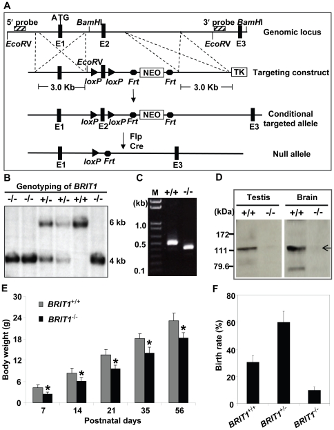
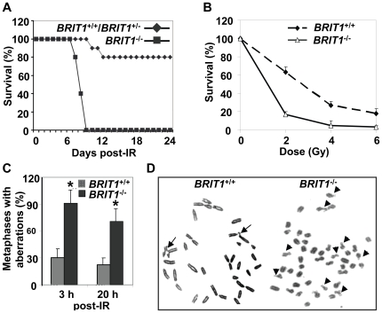
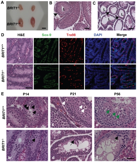
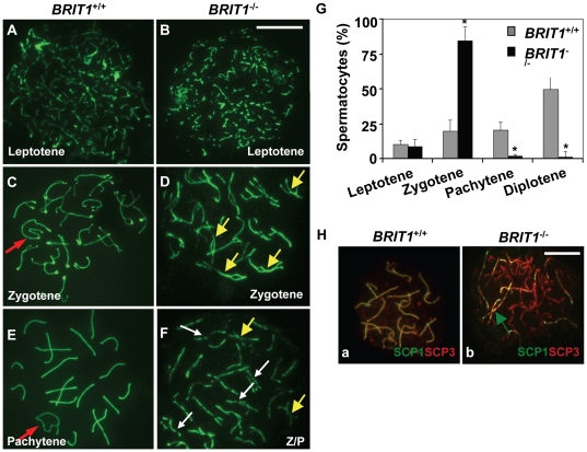
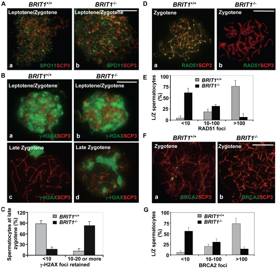
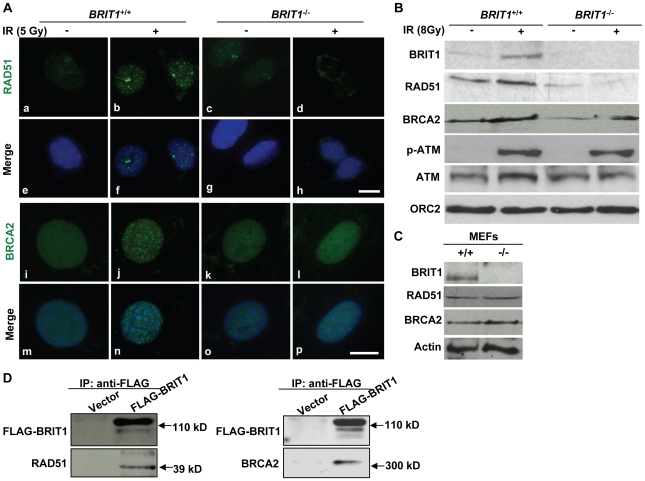
BRIT1 protein (also known as MCPH1) contains 3 BRCT domains which are conserved in BRCA1, BRCA2, and other important molecules involved in DNA damage signaling, DNA repair, and tumor suppression. BRIT1 mutations or aberrant expression are found in primary microcephaly patients as well as in cancer patients. Recent in vitro studies suggest that BRIT1/MCPH1 functions as a novel key regulator in the DNA damage response pathways. To investigate its physiological role and dissect the underlying mechanisms, we generated BRIT1(-/-) mice and identified its essential roles in mitotic and meiotic recombination DNA repair and in maintaining genomic stability. Both BRIT1(-/-) mice and mouse embryonic fibroblasts (MEFs) were hypersensitive to gamma-irradiation. BRIT1(-/-) MEFs and T lymphocytes exhibited severe chromatid breaks and reduced RAD51 foci formation after irradiation. Notably, BRIT1(-/-) mice were infertile and meiotic homologous recombination was impaired. BRIT1-deficient spermatocytes exhibited a failure of chromosomal synapsis, and meiosis was arrested at late zygotene of prophase I accompanied by apoptosis. In mutant spermatocytes, DNA double-strand breaks (DSBs) were formed, but localization of RAD51 or BRCA2 to meiotic chromosomes was severely impaired. In addition, we found that BRIT1 could bind to RAD51/BRCA2 complexes and that, in the absence of BRIT1, recruitment of RAD51 and BRCA2 to chromatin was reduced while their protein levels were not altered, indicating that BRIT1 is involved in mediating recruitment of RAD51/BRCA2 to the damage site. Collectively, our BRIT1-null mouse model demonstrates that BRIT1 is essential for maintaining genomic stability in vivo to protect the hosts from both programmed and irradiation-induced DNA damages, and its depletion causes a failure in both mitotic and meiotic recombination DNA repair via impairing RAD51/BRCA2's function and as a result leads to infertility and genomic instability in mice.


Conflict of interest statement

The authors have declared that no competing interests exist.

Figures

Figure 1
Figure 1. Generation and growth retardation of BRIT1-deficient mouse.
(A) Schematic diagram of the BRIT1 targeting strategy. The BRIT1 targeting was described in detail in Materials and Methods. The positions of 5′ and 3′ flanking probes were shown as diagonally striped boxes. E1, E2, and E3 were the first three exons of BRIT1. (B) Genotyping of BRIT1 +/+, BRIT1 +/− and BRIT1 −/− mice. Genotyping was performed by Southern blot analyses. A 6 kb fragment was detected for WT and a 4 kb fragment was for the mutant mice. (C) Disruption of BRIT1 transcript in BRIT1 −/− mice. Total testes RNA from the indicated mice were extracted and subjected to RT-PCR. The products of RT-PCR were 514 bp (WT allele) and 422 bp (mutant allele). M, DNA marker. (D) Loss of BRIT1 protein in BRIT1 −/− mice. Total proteins from the indicated tissues were extracted and subjected to Western blot using an anti-BRIT1 antibody. Arrow, the full-length BRIT1 protein. (E) BRIT1 −/− mice were growth-retarded. Body weights of BRIT1 +/+ and BRIT1 −/− littermates (n = 50 for each genotype in each age group) were measured at the postnatal day (P) 7, P14, P21, P35, and P56. *, P<0.05 compared with WT. (F) Lower birth rate of BRIT1 −/− mice. Birth rates of BRIT1 +/+, BRIT1 +/− and BRIT1 −/− mice were calculated from the offspring of self-cross of BRIT1 +/− mice (n = 400).
Figure 2
Figure 2. BRIT1-deficient mice and MEFs showed hypersensitive to γ-irradiation.
(A) BRIT1 −/− mice were hypersensitive to γ-irradiation (IR). BRIT1 +/+, BRIT1 +/− and BRIT1 −/− littermates (n = 30 for each genotype) were exposed to 7 Gy of whole-body IR, and were then monitored for 4 weeks. (B) BRIT1 −/− MEFs were more sensitive to IR. Radiosensitivity of BRIT1 +/+ and BRIT1 −/− MEFs was plotted as the fraction of surviving cells relative to unirradiated cells of the same genotype. (C,D) Enhanced chromosome aberrations in BRIT1 −/− MEFs. BRIT1 +/+ and BRIT1 −/− MEFs were γ-irradiated with 1 Gy, collected at 3 h or 20 h after IR, and subjected to metaphase spread assay. At least 30–35 metaphase spreads were analyzed for chromosome aberrations for each genotype. The main type of chromosome aberrations is chromatid breaks. Representative aberrations in BRIT1 +/+ and BRIT1 −/− MEFs at 3 h post-IR were shown in (D). Arrows represent chromatid breaks.
Figure 3
Figure 3. BRIT1-deficient male were infertile and exhibited meiotic defects.
(A) Smaller testes in BRIT1 −/− mice. Shown here are the testes from the indicated mice at P28. Scale bar, 5 mm. (B,C) Thinner seminiferous epithelia in BRIT1 −/− mice. Testes sections from BRIT1 +/+ (B) and BRIT1 −/− (C) mice at P28 were stained with haematoxylin-eosin (H&E). Arrows, the seminiferous tubules; L, lumen of the seminiferous tubules. Scale bar, 50 µm. (D) Mitosis was not defective in BRIT1 −/− spermatogonia and Sertoli cells. Testes at P7 were either stained with H&E or double-stained with anti-Tra98 and anti-Sox9 antibodies using immunofluorescent staining. Scale bar, 50 µm. (E) Meiotic dysregulation in BRIT1 −/− spermatocytes. H&E staining was performed in testis sections from BRIT1 +/+ and BRIT1 −/− littermates at P14, P21, and P56. Black arrows: spermatocytes at leptotene/zygotene (a, b, d, f); white arrows: spermatocytes at diplotene (c); green arrows: elongate spermatids (e). Scale bar, 50 µm.
Figure 4
Figure 4. Meiosis in BRIT1-deficient spermatocytes was arrested prior to the pachytene stage with aberrant chromosomal synapsis.
The staging of spermatocytes was examined in three pairs of WT and mutant mice by using anti-SCP3 immunostaining of the spermatocyte nuclei spread. (A,B) No defects in BRIT1−/− leptotene spermatocytes. The leptotene spermatocytes were detected in WT (A) and BRIT1 −/− testes (B). Scale bar, 10 µm. (C,D) Aberrant bivalents in BRIT1 −/− zygotene spermatocytes. Compared to the WT (C) with normal synapsis initiated typically at the distal ends of the acrocentric chromosomes, many mutant bivalents showed interstitial initiation of synapsis with asynapsis on either side of the contact (D). Red arrow, sex body; yellow arrows, interstitial synapsis. (E,F) BRIT1 −/− spermatocytes were prone to be fragmented prior to the pachytene stage. Unlike WT with typical pachytene morphology (E), the mutant (F) was arrested prior to the pachytene stage or zygotene/pachytene transition stage and showed fragmented chromosome (white arrows). Red arrow, sex body; yellow arrows, interstitial synapsis; Z/P represents aberrant late zygotene/pachytene transition stage; white arrows, fragmented bivalents. (G) BRIT1 −/− spermatocytes were dramatically arrested prior to the pachytene stage. Three to five hundred spermatocytes at meiosis I during the first wave were counted. Data are plotted as average percentage (mean±SD) determined from three pairs of mutant and WT mice. *, P≤0.01 (mutant versus WT). (H) Defective synapsis in BRIT1−/− spermatocytes. Synapsis here was determined by SCP1 staining. The complete bivalents were detected at zygotene/pachytene transition in WT spermatocytes (a). However, the mutant spermatocytes exhibited incomplete, dashed-line shape bivalents in many homologs during zygotene or zygotene/pachytene transition stage (b). Green arrow represents the incomplete, dashed-line shape synapsis (bivalents). Scale bar, 10 µm.
Figure 5
Figure 5. Recombination DNA repair was impaired in BRIT1-deficient spermatocytes.
BRIT1 +/+ and BRIT1 −/− spermatocytes were obtained from the indicated mice (P17.5–P22.5), and subjected for chromosome spreading and then double-stained immunofluorescently with anti-SCP3/anti-SPO11, anti-SCP3/anti-γ-H2AX, anti-SCP3/anti-RAD51, or anti-SCP3/anti-BRCA2 antibodies, respectively. Anti-SCP3 staining was used to determine the stage of each spermatocyte. Total around 400 chromosome spreads from three sets of mice were analyzed. Scale bar, 10 µm. (A) DNA double-strand breaks (DSBs) formation in meiosis is not affected by loss of Brit1. SPO11 foci had the similar pattern in WT and BRIT1-deficient spermatocytes. (B) γ-H2AX foci formation at leptotene/zygotene was comparable between WT and mutant spermatocytes. Both WT (a) and mutant (b) spermatocytes exhibited abundant γ-H2AX foci in response to DSBs at leptotene/zygotene stages, although γ-H2AX foci disappeared at the late zygotene/pachytene stage except in the XY body in WT (c), and they were sustained on asynapsed autosomal homologs in the mutant spermatocytes at late zygotene (d). (C) Retained γ-H2AX foci during late zygotene spermatocyte in BRIT1-deficient spermatocytes. Value shown here represents the mean±SD from ∼50 late zygotene spermatocytes for each genotype. (D) RAD51 foci were dramatically decreased in zygotene stage of BRIT1 −/− spermatocytes. RAD51 foci were formed intensively on zygotene homologs in WT (a) while few foci were detected in the BRIT1 −/− zygotene (b). (E) Remarkable reduction in the number of RAD51 foci per leptotene/zygotene spermatocyte in BRIT1-deficient spermatocytes. Value shown here represents the mean ± SD from ∼50 leptotene/zygotene spermatocytes for each genotype. (F) BRCA2 foci formation was disrupted in BRIT1 −/− spermatocytes. BRCA2 foci were formed intensively on bivalents at zygotene in WT (a) but not in the BRIT1 −/− zygotene (b). (G) Substantial reduction in the number of BRCA2 foci per leptotene/zygotene spermatocyte in BRIT1-deficient spermatocytes. Value shown here represents the mean ± SD from ∼50 leptotene/zygotene spermatocytes for each genotype.
Figure 6
Figure 6. BRIT1 is required for recruitment of RAD51/BRCA2 to the IR-induced DNA damage sites.
(A) RAD51/BRCA2 foci formation was inhibited in IR-treated BRIT1 −/− MEFs. The WT and mutant MEFs on the coverslips were treated with or without ionizing radiation (5 Gy), then subjected to immunofluorescent staining 30 min later. IR-induced RAD51 foci were diminished in BRIT1 −/− MEFs (d) compared to those in WT (b). Similarly, compared to WT (j), BRCA2 foci in mutant MEFs (l) were barely formed in response to IR. Scale bar, 10 µm. (B) Chromatin-bound RAD51 and BRCA2 were dramatically reduced in IR-treated BRIT1 −/− MEFs. MEFs were treated with or without ionizing radiation (8Gy) and collected 1 h later. Chromatin pellets were then subjected to chromatin isolation and Western blot analysis, and probed with antibodies against BRIT1, RAD51, BRCA2, p-ATM, ATM or ORC2, respectively. ORC2 was used as a loading control. Chromatin-bound p-ATM or ATM remained the same between the WT and the mutant cells. However, there was much fewer RAD51 or BRCA2 bound to chromatin in the mutant MEFs as compared to the WT. p-ATM: phosphorylated ATM. (C) RAD51 or BRCA2 protein expression was not altered due to loss of BRIT1. Total protein lysates from indicated MEFs were used to detect the protein levels of RAD51/BRCA2. RAD51/BRCA2 expression was comparable between the WT and mutant MEFs. (D) BRIT1 physically associated with RAD51/BRCA2. The immortalized BRIT1 +/+ MEFs were transfected with vector or FLAG-BRIT1 and the cell lysates were collected 1 h after irradiation (8Gy), subjected to anti-FLAG immunoprecipitation assay, separated by SDS-PAGE, and blotted with anti-FLAG, anti-RAD51, or anti-BRCA2 antibodies, respectively.

Similar articles

Cited by

References

    1. Pastink A, Eeken JC, Lohman PH. Genomic integrity and the repair of double-strand DNA breaks. Mutat Res. 2001;480–481:37–50. - PubMed
    1. Franco S, Alt FW, Manis JP. Pathways that suppress programmed DNA breaks from progressing to chromosomal breaks and translocations. DNA Repair. 2006;5:1030–1041. - PubMed
    1. Pandita TK, Hittelman WN. The contribution of DNA and chromosome repair deficiencies to the radiosensitivity of ataxia-telangiectasia. Radiat Res. 1992;131:214–223. - PubMed
    1. Kuzminov A. Collapse and repair of replication forks in Escherichia coli. Mol Microbiol. 1995;16:373–384. - PubMed
    1. Richardson C, Horikoshi N, Pandita TK. The role of the DNA double-strand break response network in meiosis. DNA Repair. 2004;3:1149–1164. - PubMed

Publication types