

PAS-positive lymphocyte vacuoles can be used as diagnostic screening test for Pompe disease
- PMID: 20107902
- PMCID: PMC2861182
- DOI: 10.1007/s10545-009-9027-4
PAS-positive lymphocyte vacuoles can be used as diagnostic screening test for Pompe disease
Abstract
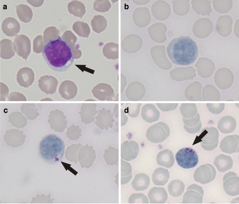
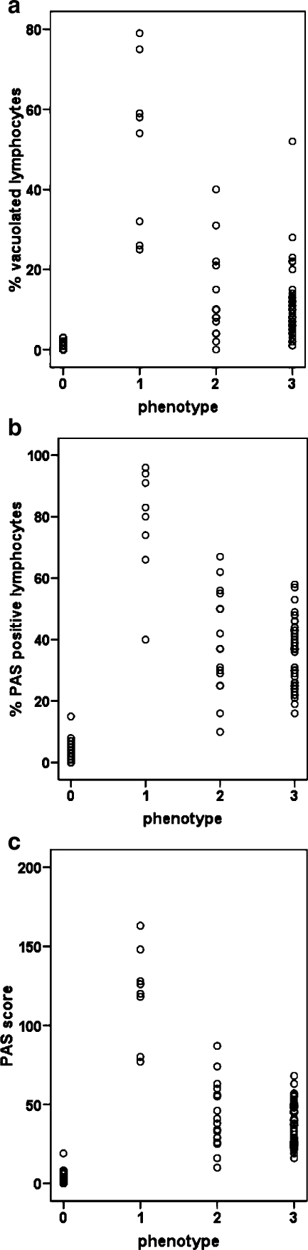
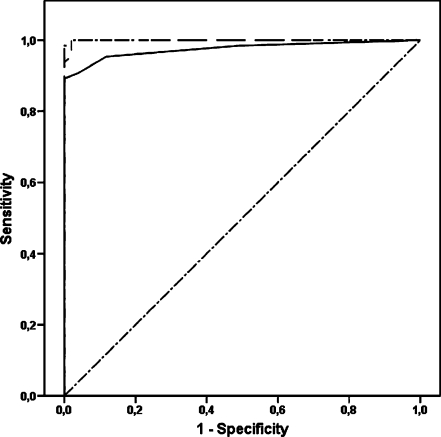
Screening of blood films for the presence of periodic acid-Schiff (PAS)-positive lymphocyte vacuoles is sometimes used to support the diagnosis of Pompe disease, but the actual diagnostic value is still unknown. We collected peripheral blood films from 65 untreated Pompe patients and 51 controls. Lymphocyte vacuolization was quantified using three methods: percentage vacuolated lymphocytes, percentage PAS-positive lymphocytes, and a PAS score depending on staining intensity. Diagnostic accuracy of the tests was assessed using receiver operating characteristic (ROC) curves. All three methods fully discerned classic infantile patients from controls. The mean values of patients with milder forms of Pompe disease were significantly higher than those of controls, but full separation was not obtained. The area under the ROC curve was 0.98 for the percentage vacuolated lymphocytes (optimal cutoff value 3; sensitivity 91%, specificity 96%) and 0.99 for the percentage PAS-positive lymphocytes and PAS score (optimal cutoff value 9; sensitivity 100%, specificity 98%). Our data indicate that PAS-stained blood films can be used as a reliable screening tool to support a diagnosis of Pompe disease. The percentage of PAS-positive lymphocytes is convenient for use in clinical practice but should always be interpreted in combination with other clinical and laboratory parameters.
Figures
Similar articles
-
Vacuolated PAS-Positive Lymphocytes on Blood Smear: An Easy Screening Tool and a Possible Biomarker for Monitoring Therapeutic Responses in Late Onset Pompe Disease (LOPD).Front Neurol. 2018 Oct 22;9:880. doi: 10.3389/fneur.2018.00880. eCollection 2018. Front Neurol. 2018. PMID: 30405515 Free PMC article.
-
Vacuolated PAS-positive lymphocytes as an hallmark of Pompe disease and other myopathies related to impaired autophagy.J Cell Physiol. 2018 Aug;233(8):5829-5837. doi: 10.1002/jcp.26365. Epub 2018 Feb 22. J Cell Physiol. 2018. PMID: 29215735
-
Quantification of muscle pathology in infantile Pompe disease.Neuromuscul Disord. 2017 Feb;27(2):141-152. doi: 10.1016/j.nmd.2016.10.010. Epub 2016 Nov 3. Neuromuscul Disord. 2017. PMID: 27927596
-
Pompe disease: early diagnosis and early treatment make a difference.Pediatr Neonatol. 2013 Aug;54(4):219-27. doi: 10.1016/j.pedneo.2013.03.009. Epub 2013 Apr 28. Pediatr Neonatol. 2013. PMID: 23632029 Review.
-
The clinical and electrodiagnostic characteristics of Pompe disease with post-enzyme replacement therapy findings.Clin Neurophysiol. 2011 Nov;122(11):2312-7. doi: 10.1016/j.clinph.2011.04.016. Epub 2011 May 13. Clin Neurophysiol. 2011. PMID: 21570905 Review.
Cited by
-
Vacuolated PAS-Positive Lymphocytes on Blood Smear: An Easy Screening Tool and a Possible Biomarker for Monitoring Therapeutic Responses in Late Onset Pompe Disease (LOPD).Front Neurol. 2018 Oct 22;9:880. doi: 10.3389/fneur.2018.00880. eCollection 2018. Front Neurol. 2018. PMID: 30405515 Free PMC article.
-
Effects of cadmium exposure on antioxidant enzymes and histological changes in the mud shrimp Austinogebia edulis (Crustacea: Decapoda).Environ Sci Pollut Res Int. 2019 Mar;26(8):7752-7762. doi: 10.1007/s11356-018-04113-x. Epub 2019 Jan 23. Environ Sci Pollut Res Int. 2019. PMID: 30673948
-
Detecting glycogen in peripheral blood mononuclear cells with periodic acid schiff staining.J Vis Exp. 2014 Dec 23;(94):52199. doi: 10.3791/52199. J Vis Exp. 2014. PMID: 25548935 Free PMC article.
-
Hematological Findings in Lysosomal Storage Disorders: A Perspective from the Medical Laboratory.EJIFCC. 2022 Apr 11;33(1):28-42. eCollection 2022 Apr. EJIFCC. 2022. PMID: 35645695 Free PMC article.
-
Diagnostic tools in late onset Pompe disease (LOPD).Ann Transl Med. 2019 Jul;7(13):286. doi: 10.21037/atm.2019.06.60. Ann Transl Med. 2019. PMID: 31392198 Free PMC article. Review.
References
-
- Bain JB. Blood cells a practical guide. 2. Oxford: Blackwell Science; 1995. pp. 186–188.
-
- de Barsy T, Hers HG. Biochemical and ultrastructural study of leucocytes in type II glycogenosis. Arch Int Physiol Biochim. 1975;83:954–955. - PubMed
-
- Hayhoe FGJ, Quaglino D. Carbohydrates. Haematological cytochemistry. New York: Churchill Livingstone; 1980. pp. 50–67.
-
- Hirschhorn R, Reuser AJJ (2000) Glycogen Storage Disease type II; acid α-Glucosidase (Acid Maltase) deficiency. In: Scriver CR, Beaudet AL, Sly W, Valle D (eds) 8th edn, vol III. Mc Graw-Hill, New York, pp 3389–3420
Publication types
MeSH terms
Substances
LinkOut - more resources
Full Text Sources
Medical
