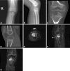
Case report: primary aneurysmal bone cyst of the epiphysis
- PMID: 20107940
- PMCID: PMC2835586
- DOI: 10.1007/s11999-010-1228-5
Case report: primary aneurysmal bone cyst of the epiphysis
Abstract
Aneurysmal bone cysts are benign active or aggressive bone tumors that commonly arise in the long bones, especially the femur, tibia, and humerus and the posterior elements of the spine. Aneurysmal bone cysts affect all age groups but are more common before skeletal maturity (first two decades of life). They usually involve the metaphysis or metadiaphyseal region of long bones. Although juxtaphyseal lesions abutting the growth plate and extending into the epiphysis have been described, there is no report of an aneurysmal bone cyst entirely and primarily located in the epiphysis. We report on a 3-year-old boy who presented with an entirely contained aneurysmal bone cyst to the proximal tibial epiphysis. We discuss the clinical presentation, diagnosis, including imaging and pathology, and treatment. A review of the pertinent literature also is presented.
Figures


Similar articles
-
Primary aneurysmal bone cyst of the distal tibial epiphysis: a case report.J Pediatr Orthop B. 2014 May;23(3):266-9. doi: 10.1097/BPB.0000000000000014. J Pediatr Orthop B. 2014. PMID: 24217294
-
Juxtaphyseal aneurysmal bone cysts.Clin Orthop Relat Res. 1999 Jul;(364):205-12. doi: 10.1097/00003086-199907000-00026. Clin Orthop Relat Res. 1999. PMID: 10416410
-
Aneurysmal bone cyst arising from a fibrous metaphyseal defect in a toddler.Clin Orthop Relat Res. 2002 Feb;(395):216-20. doi: 10.1097/00003086-200202000-00025. Clin Orthop Relat Res. 2002. PMID: 11937884
-
Chondroblastoma of the patella with secondary aneurysmal bone cyst, an easily misdiagnosed bone tumor:a case report with literature review.BMC Musculoskelet Disord. 2021 Apr 23;22(1):381. doi: 10.1186/s12891-021-04262-0. BMC Musculoskelet Disord. 2021. PMID: 33892701 Free PMC article. Review.
-
Aggressive aneurysmal bone cyst of the proximal humerus. A case report.Clin Orthop Relat Res. 2000 Jan;(370):212-8. doi: 10.1097/00003086-200001000-00021. Clin Orthop Relat Res. 2000. PMID: 10660716 Review.
Cited by
-
Aneurysmal Bone Cyst-An Unusual Presentation of Wrist Pain.J Wrist Surg. 2019 Feb;8(1):76-79. doi: 10.1055/s-0038-1660810. Epub 2018 Jun 13. J Wrist Surg. 2019. PMID: 30723607 Free PMC article.
-
Case reports with literature review of an aneurysmal bone cyst in the maxilla and mandible of two juvenile dogs.Front Vet Sci. 2025 Jul 31;12:1632403. doi: 10.3389/fvets.2025.1632403. eCollection 2025. Front Vet Sci. 2025. PMID: 40822658 Free PMC article.
-
Pelvic Aneurysmal Bone Cyst in an Adolescent: A Case Report and Literature Review.Cureus. 2020 Aug 3;12(8):e9534. doi: 10.7759/cureus.9534. Cureus. 2020. PMID: 32775117 Free PMC article.
-
Aneurysmal bone cyst of the orbit : a case report with literature review.J Korean Neurosurg Soc. 2012 Feb;51(2):113-6. doi: 10.3340/jkns.2012.51.2.113. Epub 2012 Feb 29. J Korean Neurosurg Soc. 2012. PMID: 22500206 Free PMC article.
-
Joint Reconstruction using Tricortical Iliac Crest Bone Graft Block for Intra-Articular Extension of Aneurysmal Bone Cyst of Distal Tibia in a Skeletally Mature Patient - A Case Report and Review of Literature.J Orthop Case Rep. 2025 Mar;15(3):37-42. doi: 10.13107/jocr.2025.v15.i03.5326. J Orthop Case Rep. 2025. PMID: 40092234 Free PMC article.
References
-
- Bollini G, Jouve JL, Cottalorda J, Petit P, Panuel M, Jacquemier M. Aneurysmal bone cyst in children: analysis of twenty-seven patients. J Pediatr Orthop B. 1998;7:274–285. - PubMed
-
- Capanna R, Bettelli G, Biagini R, Ruggieri P, Bertoni F, Campanacci M. Aneurysmal cysts of long bones. Ital J Orthop Traumatol. 1985;11:409–417. - PubMed
-
- Capanna R, Sudanese A, Baldini N, Campanacci M. Phenol as an adjuvant in the control of local recurrence of benign neoplasms of bone treated by curettage. Ital J Orthop Traumatol. 1985;11:381–388. - PubMed
Publication types
MeSH terms
LinkOut - more resources
Full Text Sources

