. 2010 Aug;28(8):1000-6.
doi: 10.1002/jor.21087.

Rejuvenation of the inflammatory system stimulates fracture repair in aged mice

Affiliations

Rejuvenation of the inflammatory system stimulates fracture repair in aged mice

Zhiqing Xing et al. J Orthop Res. 2010 Aug.

Abstract

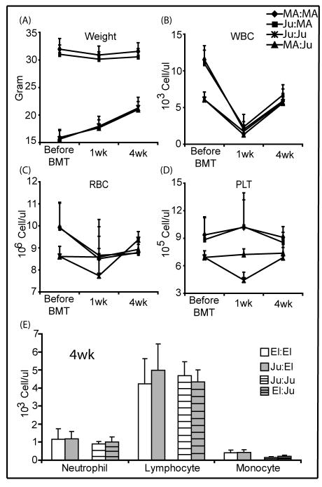
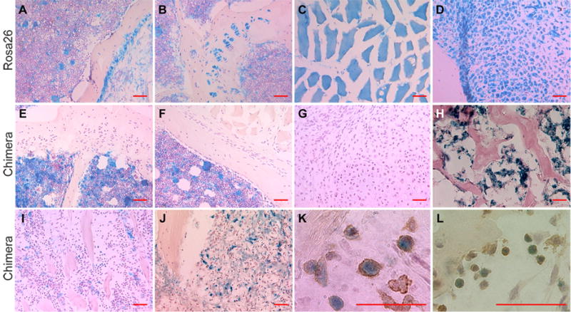
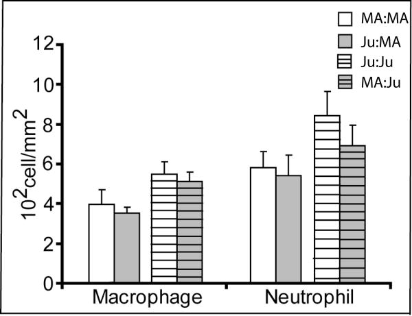
Age significantly reduces the regenerative capacity of the skeleton, but the underlying causes are unknown. Here, we tested whether the functional status of inflammatory cells contributes to delayed healing in aged animals. We created chimeric mice by bone marrow transplantation after lethal irradiation. In this model, chondrocytes and osteoblasts in the regenerate are derived exclusively from host cells while inflammatory cells are derived from the donor. Using this model, the inflammatory system of middle-aged mice (12 month old) was replaced by transplanted bone marrow from juvenile mice (4 weeks old), or age-matched controls. We found that the middle-aged mice receiving juvenile bone marrow had larger calluses and more bone formation during early stages and faster callus remodeling at late stages of fracture healing, indicating that inflammatory cells derived from the juvenile bone marrow accelerated bone repair in the middle-aged animals. In contrast, transplanting bone marrow from middle-aged mice to juvenile mice did not alter the process of fracture healing in juvenile mice. Thus, the roles of inflammatory cells in fracture healing may be age-related, suggesting the possibility of enhancing fracture healing in aged animals by manipulating the inflammatory system.


Figures

Figure 1
Figure 1. Recovery from irradiation and bone marrow transplantation (BMT)
(A) 4 weeks after BMT, there was no difference in weight, (B) white blood cell (WBC), (C) red blood cell (RBC), (D) platelet (PLT), and (E) differential counts of white blood cells among the chimeric and control groups (MA:MA, n = 22 at each time point; Ju:MA mice, n = 21 before BMT, n = 19 at 1wk or 4wk; Ju:Ju and MA:Ju, n=8/time point).
Figure 2
Figure 2. Distribution of donor bone marrow derived cells in the chimeras
(A) Cells in bone marrow, (B) periosteum, growth plate, (C) muscle fibers, and (D) chondrocytes in the fracture callus were stained blue by X-gal staining in ROSA26 mice. (E) In chimeric mice, donor cells were not observed in growth plate, (F) cortical bone, periosteum, muscle, and (G) cartilage and (H) bone in the callus. (I) 7 days after fracture donor cells (blue) were present at the fracture site and (J) in granulation tissue. (K) Donor cells were stained with F4/80 or (L) MCA771G. (Scale bar = 50μm)
Figure 3
Figure 3. Recruitment of inflammatory cells to fracture site
7 days after fracture, there was no difference in the densities of macrophages and neutrophils in fracture callus between Ju:MA and MA:MA as well as between MA:Ju and Ju:Ju. However, the cell densities were significantly higher in juvenile hosts than middle-aged hosts regardless of the ages of donors (p<0.001 for both cell types; n = 6 each chimeric group)
Figure 4
Figure 4. Plasma levels of IL-6 and TNF-alpha in mice with bone fracture
At day 7 after fracture, middle-aged mice (MA, n=6) had significantly higher plasma IL-6 than juvenile mice (Ju, n=6, p<0.01). After rejuvenation of bone marrow, plasma IL-6 in middle-aged mice (Ju:MA, n=6) was significantly lower than in controls (MA:MA, n=7, p=0.014). Plasma IL-6 in juvenile mice with aged marrow (MA:Ju, n=4) was higher than controls (Ju:Ju, n=5), but not significant. TNF-alpha exhibited the same trend (* p>0.05)
Figure 5
Figure 5. Histomorphometric analysis of fracture callus in chimeric mice
(A) Callus size and (B) volume of new bone were significantly larger in Ju:MA chimeras at day 7, and were significantly decreased at day 28 compared to MA:MA mice. (C) There was no difference in the volume of cartilage between these groups at day 7 and day14. At day 28, 5/7 MA:MA mice had cartilage in the callus. (* p<0.05). (D) There was no difference in Callus size, (E) volume of new bone, and (F) new cartilage between Ju:Ju and MA:Ju mice at day 7 (Ju:Ju, n=8, MA:Ju,n=8), day14 (Ju:Ju, n=7, MA:Ju, n=6), and day21 (Ju:Ju, n=7, MA:MA, n=7) after fracture.

References

    1. Einhorn TA. Enhancement of fracture-healing. J Bone Joint Surg Am. 1995;77:940–56. - PubMed
    1. Meyer RA, Meyer MH, Phieffer LS, Banks DM. Delayed union of femoral fractures in older rats:decreased gene expression. BMC Musculoskelet Disord. 2001;2:2. - PMC - PubMed
    1. Meyer J, Ralph A, Tsahakis PJ, Martin DF, et al. Age and ovariectomy impair both the normalization of mechanical properties and the accretion of mineral by the fracture callus in rats. Journal of Orthopaedic Research. 2001;19:428–435. - PubMed
    1. Meyer RA, Jr, Meyer MH, Tenholder M, et al. Gene expression in older rats with delayed union of femoral fractures. J Bone Joint Surg Am. 2003;85-A:1243–54. - PubMed
    1. Desai BJ, Meyer MH, Porter S, et al. The effect of age on gene expression in adult and juvenile rats following femoral fracture. J Orthop Trauma. 2003;17:689–98. - PubMed

Publication types