. 2010 Mar;239(3):998-1009.
doi: 10.1002/dvdy.22229.

Nesprins, but not sun proteins, switch isoforms at the nuclear envelope during muscle development

Affiliations

Nesprins, but not sun proteins, switch isoforms at the nuclear envelope during muscle development

K Natalie Randles et al. Dev Dyn. 2010 Mar.

Abstract

Nesprins are a family of nuclear transmembrane proteins anchored via Sun proteins to the nuclear membrane. Analysis of nesprins during human muscle development revealed an increase in nesprin-1-giant during early myogenesis in vitro. During the transition from immature to mature muscle fibres in vivo, nesprin-2 partly replaced nesprin-1 at the nuclear envelope and short nesprin isoforms became dominant. Sun1 and Sun2 proteins remained unchanged during this fibre maturation. In emerin-negative skin fibroblasts, nesprin-2-giant was relocated from the nuclear envelope to the cytoplasm, not to the endoplasmic reticulum, while nesprin-1 remained at the nuclear envelope. In emerin-negative keratinocytes lacking nesprin-1, nesprin-2 remained at the nuclear envelope. HeLa cell nuclear envelopes lacked nesprin-1, which was the dominant form in myoblasts, while a novel 130-kD nesprin-2 isoform dominated Ntera-2 cells. The results suggest the possibility of isoform-specific and tissue-specific roles for nesprins in nuclear positioning.


Figures

Figure 1
Figure 1
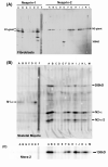
Expression of nesprin isoforms in muscle and fibroblasts. (A) Western blots of normal adult human muscle (Mus), human primary skeletal muscle cell culture (Myo) and human primary dermal fibroblasts (Fib) were probed with MANNES1A or MANNES2A antibodies (1:100) as described in Methods. The blot was reprobed with MANLAC1 mAb against lamin A/C (Manilal et al, 2004) as a loading control. Mr (relative molecular mass) markers were EZ-Run (Fisher Scientific, Loughborough, UK). (B) A second muscle biopsy developed with MANNES2A had less degradation products, though the nesprin-2-giant band was less clear. Minor bands at 180kD and 140kD may be degradation products. The gel was run as a single lane and the blot was cut into strips. MANDYS1 (1:500) to dystrophin (Nguyen thi Man et al, 1990) was used as a high Mr marker (427kD) and as an indicator of the extent of degradation of “spectrin-like” proteins in the samples. Sigma prestained Mr markers were used. (C) Mouse embryonic fibroblasts (MEFs) from lmna -/- knockout mice also express mainly nesprin-1-giant. The other bands of lower Mr do not correspond in size to any known shorter isoform. Mr markers were EZ-Run (Fisher).
Figure 2
Figure 2
Multi-epitope mAb panels confirm the identity of authentic nesprin proteins. Extracts of (A) human dermal fibroblasts, (B) adult human skeletal muscle or (C) human Ntera-2 cells, were subjected to SDS-PAGE as a strip and the western blots were developed with multiple nesprin mAbs (1:100 dilution) using a miniblotter. The mAbs vary in strength (see also Table 1), but the strongest are strong for all nesprin isoforms. In (A), MANNES2E recognizes a 300kD cross-reacting protein, similar only in size to the 300kD band in (B), which is recognized by many mAbs.
Figure 3
Figure 3
Human skin fibroblasts without emerin show normal nuclear nesprin-1 and Sun protein staining, but nuclear nesprin-2 is absent. Primary skin fibroblast cultures from a female carrier of X-linked Emery-Dreifuss MD contain both emerin-positive and emerin-negative cells as a result of random X-inactivation. Acetone-methanol-fixed cells were double-labelled with rabbit anti-emerin serum (1:100: green with a DAPI counterstain; Holt et al, 2003) and either MANNES1A (A-B, nesprin-1: red) or MANNES2A (C-F, nesprin-2: red). Emerin-negative nuclei (white arrows) had normal nuclear nesprin-1, but nuclear nesprin-2 was absent. Sun1 (H: green) was present at the nuclear rim in all cells and was not significantly different in cells lacking emerin (G: red). MEFs (mouse embryonic fibroblasts) from lmna -/- mice illustrate the redistribution of both emerin (I) and nesprin-1 (J) into the ER in the absence of lamin A/C. The emerin gene mutation was a premature stop codon in exon 4. Bars (white) = 50μm
Figure 4
Figure 4
Nesprin-2 is absent from the nuclear rim of emerin-negative skin fibroblasts from an X-EDMD patient, but western blots show that nesprin-2 levels are unchanged. (A) Staining as in Fig. 3 with single antibody labelling (green) for emerin, lamin A/C or nesprins and DAPI counterstain (blue). This patient had a frameshift deletion of bp 329-388 in the emerin gene leading to a stop after 19 amino-acids and was unrelated to the carrier in Fig. 3. Bar (white) = 20μm. (B) Monolayer cultures of control (Coriell cell line No. 8333) and X-EDMD fibroblasts were harvested with trypsin-EDTA and extracted. Extracts were subjected to SDS-PAGE on 3-15% gradient gels and blots developed with MANNES2A for nesprin-2, MANLAC1 for lamin A and MANEM5 (Manilal et al, 1996) for emerin. The lamin A band was used as a loading control and Mr markers were EZ-Run.
Figure 5
Figure 5
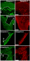
Nesprin-2 is present in emerin-negative keratinocytes in a carrier EDMD skin biopsy. Frozen sections (5 μm) were fixed with acetone-methanol and double-labelled for emerin (green) and nesprin-1 or nesprin-2 (red). Nesprin-1 is positive at the nuclear rim in dermal fibroblasts and negative in epidermal keratinocytes, both with and without emerin. White lines show the boundary between dermis (D) and epidermis (E). Unlike cultured dermal fibroblasts, all epidermal keratinocytes, with or without emerin, are positive for nesprin-2 at the nuclear rim. Bars (white) = 50μm
Figure 6
Figure 6
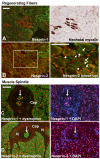
A nesprin isoform switch between immature and mature muscle fibres. Double-labels of nesprin mAbs with a polyclonal anti-dystrophin to show muscle fibre outlines were counterstained with DAPI to show nuclei. Serial sections. Bars (white) = 100μm (A, B) In a Becker MD patient, dystrophin staining is weak except in a few revertant fibres. Groups of immature, regenerating fibres are common in Becker MD and their nuclei are more brightly stained for nesprin-1 than surrounding mature fibres (A), but they have less nesprin-2 than surrounding mature fibres (B: white arrows). Immature fibres were identified using a neonatal-specific anti-myosin mAb (NCL-MHCn, Leica, Milton Keynes, UK) on a serial section. (C, D) In a child with congenital myotonic dystrophy, fibres are smaller with large central nuclei, but dystrophin staining at the sarcolemma is normal. The muscle spindle is a specialized sensory structure in muscle that contains a group of immature muscle fibres (white arrows). These are strongly positive for nesprin-1 (C) but virtually negative for nesprin-2 (D). “N” = nerve, “Cap” = external capsule of the spindle.
Figure 7
Figure 7
Levels of both Sun proteins reflect total nesprin levels at the NE in muscle, but are not dependent on whether nesprin-1 or nesprin-2 is expressed. In contrast to Fig 6AB, Sun protein levels at the NE were similar in mature and regenerating fibres (A, B). Small regenerating fibres have central nuclei, but two comparable peripheral nuclei are visible (white arrows). In a muscle spindle, the muscle fibre nuclei (inner white circle) are positive for both nesprin-1 and Sun proteins, while nuclei in the capsule (outer circle, DAPI not shown; cf Fig. 6CD) are weak or negative for both. These results were obtained with both Sun1 and Sun2 though only one is illustrated. Bar (white) = 100μm
Figure 8
Figure 8
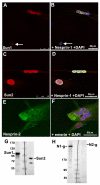
Differentiating human myoblast cultures have nesprin-1 and both Sun proteins at the nuclear envelope, but very little nesprin-2. Myoblast cultures undergoing differentiation were double-stained with anti-Sun1 serum (A: red) and MANNES1A (B: green with DAPI), or with anti-Sun2 serum (C: red) and MANNES2A (D: green with DAPI). Multinucleate myotube nuclear envelopes were always strongly positive for nesprin-1 and both Sun proteins, but mononucleated myoblasts were variable; sometimes very weak for both nesprin-1 and Sun protein (white arrows in A, B) and sometimes very strong for both (C, D). These results were obtained with both Sun1 and Sun2 though only one is illustrated. Nesprin-2 staining at the NE, however, was very weak, even in multinucleated myotubes (E: MANNES2A, green, F: anti-emerin serum, red with DAPI). Western blots confirm the presence of both Sun proteins (G) and confirm the increase in nesprin-1, but not nesprin-2, in myotubes (H), relative to myoblasts in Fig. 1A.
Figure 9
Figure 9
Nesprin-2 is cytoplasmic or perinuclear in HeLa and in Ntera-2 cells, which express a novel 130kD form. In HeLa cells, (a) nesprin-1 is absent and (b) nesprin-2 is perinuclear in many cells, though some show nuclear rim staining. (c) In the Ntera-2 human neurogenic cell line, nesprin-2 is sometimes perinuclear and close to the Golgi apparatus, but does not colocalize with it. The inset shows that many Ntera-2 nuclei have more typical nuclear rim staining for nesprin-2. Cells fixed with acetone-methanol were used for double-label with rabbit anti-GM130 antibody and either MANNES1A or MANNES2A mouse mAb (1:3 dilution). Bars (white) = 25μm For the western blot, all cell pellets were extracted under conditions designed to minimize proteolysis. Dermal fibroblasts (Fib) have a single band at 800kD with only traces of possible degradation products. Ntera cells have a clear main band at 130kD that cannot be attributed to degradation, but corresponds to no known isoform. HeLa cells also produce lower Mr nesprins (a separate blot was aligned using the EZ-run Mr markers).

Similar articles

Cited by

References

    1. Apel ED, Lewis RM, Grady RM, Sanes JR. Syne-1, a dystrophin- and Klarsicht-related protein associated with synaptic nuclei at the neuromuscular junction. J. Biol. Chem. 2000;275:31986–31995. - PubMed
    1. Barbet JP, Thornell LE, Butler-Browne GS. Immunocytochemical characterisation of two generations of fibers during the development of the human quadriceps muscle. Mech Dev. 1991;35:3–11. - PubMed
    1. Crisp M, Liu Q, Roux K, Rattner JB, Shanahan C, Burke B, Stahl PD, Hodzic D. Coupling of the nucleus and cytoplasm: role of the LINC complex. J. Cell Biol. 2006;172:41–53. - PMC - PubMed
    1. Furling D, Lam LT, Agbulut O, Butler-Browne GS, Morris GE. Changes in myotonic dystrophy protein kinase levels and muscle development in congenital myotonic dystrophy. Am J Pathol. 2003;162:1001–1009. - PMC - PubMed
    1. Gough LL, Fan J, Chu S, Winnick S, Beck KA. Golgi Localization of Syne-1. Mol. Biol. Cell. 2003;14:2410–2424. - PMC - PubMed

Publication types

MeSH terms