

Recent conclusions regarding the reconstructive microsurgery of peripheral nerves
- PMID: 20108464
- PMCID: PMC5654069
Recent conclusions regarding the reconstructive microsurgery of peripheral nerves
Abstract
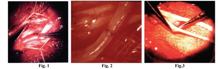
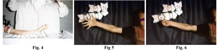
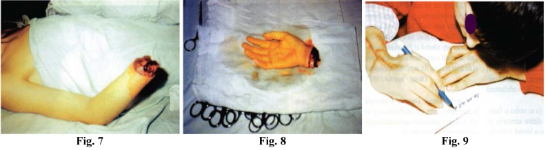
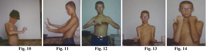
The introducing of reconstructive microsurgery has meant not only the addition of microsurgical microscopes and instruments, but a change, a progress towards a new concept, the concept of the microsurgical reconstruction of tissues. The microscope and the instruments themselves are only a means of utilizing this new concept to good effect since the mere use of the microscope and of the instruments according to the old concept of tissue reconstruction cannot be considered to be reconstructive microsurgery. From December 1979 through to December 2005, more than 3000 patients with peripheral nerve lesions were operated on by the same microsurgeon, the author Doina Ionescu-Dumitrescu. The conclusions are based on the following: A huge amount of work involved in carrying out microsurgical reconstructions of over 7500 peripheral nerves in over 3000 patients, 1800 of which were nerve transplants for defects of peripheral nerves of the extremities, for posttraumatic brachial plexus paralyses (91), for replantations and/or revascularizations following partial or complete amputations of the extremities (24 out of which 23 successful) or for free transfers of functional composite tissues (53). For a more accurate picture of such an effort one should consider the operation time that these types of reconstruction involve: between 3 and 12 hours for each patient under general anaesthesia and for both the anaesthetist and the microsurgeon. Experimental microsurgery on rabbit ears The results of the histopathological examination of 500 postoperative neuromas of peripheral nerves repaired traditionally. The Moberg test. Pre, intra and postoperative monthly observations of the patients until their full recovery according to the criteria set by the International Reconstructive Microsurgery Society (postoperative intervals of 6-12-24 months). Taking pictures and recording pre, intra and postoperative stages. The patients' professional, social and familial reintegration. The patients' state of mind; level of cooperation. Comparing results with those of classic and palliative repairs. Comparing the data resulting from this experience with the information provided by the specialist literature of the world. Completing the internationally defined reconstructive procedures with the personal ones, to produce a new concept.
Figures
Similar articles
-
[Microsurgical reconstructive-rehabilitative operations in traumatic lesions of the peripheral nerves].Zh Vopr Neirokhir Im N N Burdenko. 1989 Nov-Dec;(6):15-6. Zh Vopr Neirokhir Im N N Burdenko. 1989. PMID: 2629431 Russian.
-
[The use of the fibrin glue in the peripheral nerves reconstructions].Polim Med. 2006;36(2):11-5. Polim Med. 2006. PMID: 17022152 Polish.
-
[The surgery of peripheral nerves. New perspectives in treatment by the microsurgical techniques (author's transl)].Acta Chir Belg. 1979 Jan-Feb;78(1):47-56. Acta Chir Belg. 1979. PMID: 433509 French.
-
Microsurgical Tissue Transfer in Complex Upper Extremity Trauma.Clin Plast Surg. 2020 Oct;47(4):521-534. doi: 10.1016/j.cps.2020.06.013. Epub 2020 Aug 12. Clin Plast Surg. 2020. PMID: 32892798 Review.
-
Nerve reconstruction in the hand and upper extremity.Clin Plast Surg. 2011 Oct;38(4):643-60. doi: 10.1016/j.cps.2011.07.008. Clin Plast Surg. 2011. PMID: 22032591 Review.
References
-
- Terzis Julia K. Microreconstruction of Nerve Injuries. W.B. Saunders Company; 1987.
-
- Terzis Julia, Smith Kevin L. The Peripheral Nerve Structure, Function and Reconstruction. New York: Raven Press; 1990.
-
- Terzis Julia K. Microreconstruction of Nerve Injuries. W.B. Saunders Company; 1987.
-
- Rollin Daniel, Julia K. Terzis. Reconstructive Microsurgery. Little, Brown and Company; 1977.
-
- Jewett Don L, McCarroll H. Relton. Nerve Repair and Regeneration. The Mosby Company; 1980.
MeSH terms
LinkOut - more resources
Full Text Sources
Medical