Review
. 2009 Jan-Mar;2(1):5-17.

Genetics of craniosynostosis: review of the literature

Affiliations
Review

Genetics of craniosynostosis: review of the literature

Alexandru Vlad Ciurea et al. J Med Life. 2009 Jan-Mar.

Abstract

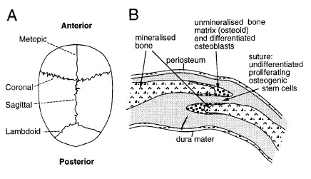
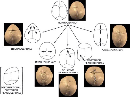
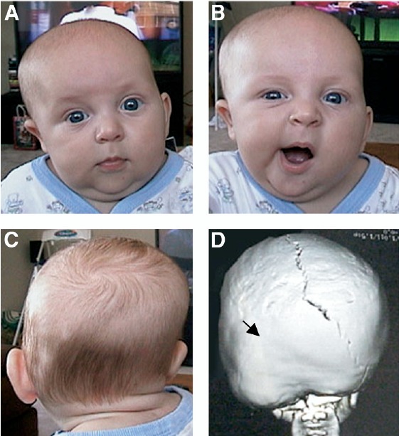
Craniosynostosis represents a defection of the skull caused by early fusion of one or more cranial sutures. The shape alteration of the cranial vault varies, depending on the fused sutures, so that compensatory growth occurs in dimensions not restricted by sutures. Craniosynostosis can be divided into two main groups: syndromic and nonsyndromic. Nonsyndromic craniosynostosis is typically an isolated finding that is classified according to the suture(s) involved. Syndromic craniosynostosis is associated with various dysmorphisms involving the face, skeleton, nervous system and is usually accompanied by developmental delay. In the last 15 years, research on craniosynostosis has progressed from the description of gross abnormalities to the understanding of the genetic basis of certain cranial deformities. Mutations in the genes encoding fibroblast growth factor receptors 1, 2 and 3 (FGFR-1, FGFR-2, FGFR-3), TWIST and MSX2 (muscle segment homebox 2) have been identified in certain syndromic craniosynostosis. The molecular basis of many types of syndromic craniosynostosis is known and diagnostic testing strategies will often lead to a specific diagnosis. Although the clarification of a genetic lesion does not have a direct impact on the management of the patient in many cases, there is a significant benefit in providing accurate prenatal diagnosis. This review summarizes the available knowledge on cranisynostosis and presents a graduated strategy for the genetic diagnosis of these craniofacial defects.


Figures

Fig. 1
Fig. 1
From De Humani Corporis Fabrica, Andreas Vesalius, 1543. Skulls depicting absence of cranial sutures and abnormal skull shapes. Reproducted with permission of []: Cunningham et al.: Syndromic craniosynostosis: from history to hydrogen bonds, Orthod Craniofacial Res 10, 2007; 67–81.
Fig. 2
Fig. 2
Normal cranial suture development. (A) View of child’s skull from above, showing position of the major sutures. Coronal craniosynostosis leads to a short, broad skull; conversely, sagittal synostosis leads to a long, narrow skull. (B) Diagrammatic cross section through coronal suture. The skull bones overlap slightly. In craniosynostosis, the narrow space separating the bones is obliterated. Reproducted with permission from [] Wilkie AOM: Craniosynostosis: genes and mechanisms, Human Molecular Genetics, 1997, Vol. 6, No. 10 Review.
Fig. 3
Fig. 3
Schematic presentation of the calvarial sutures and the skull deformities resulting from the synostosis of a particular suture. The arrows indicate the direction of skull growth. 3D-CT images of patients illustrate types of craniosynotosis. Reproducted with permission from [] Boyadjiev SA: Genetic analysis of non-syndromic craniosynostosis, Orthod Craniofacial Res 10, 2007; 129–137).
Fig. 4
Fig. 4
Posterior plagiocephaly caused by lambdoidal synostosis. (A and B) Frontal and (C) posterior views of an infant with left lambdoidal synostosis; the 3D-CT reconstruction (D, posterior view) shows premature fusion of the left lambdoidal suture (arrow). Note the facial asymmetry of the patient secondary to the lambdoidal syostosis (panel B) that does not occur in positional plagiocephaly. Reproducted with permission from: Kimonis Virginia, Gold JA, Hoffman T, Panchal J, Boyadjiev S: Genetics of Craniosynostosis, Semin Pediatr Neurol 14:150-161, 2007. [].
Fig. 5
Fig. 5
Classic features of Apert syndrome including turribrachycephaly, proptosis, midface hypoplasia and syndactyly. Airway compromise, because of midface hypoplasia, necessitated tracheostomy. Reproducted with permission from Cunningham et al. []
Fig. 6
Fig. 6
Original cases described by Crouzon demonstrating prognathism, maxillary hypoplasia, exophthalmos, papilledema, and divergent strabismus. Reproducted with permission from Cunningham et al. []
Fig. 7
Fig. 7
Original family with autosomal dominant coronal synostosis reported by Glass et al. Note the mild midfacial hypoplasia, hypertelorism, and downslanting palpebral fissures. This family later found to harbor the FGFR (3P250R) mutation of Muenke syndrome.Reproducted with permission from Cunningham et al. []
Fig. 8
Fig. 8
Craniofacial and skin findings in Crouzonodermoskeletal syndrome. After successful cranioplasty to treat Kleeblatschaedel skull deformity, he has residual midfacial hypoplasia. Note periorbital and perioral acanthosis nigricans. This patient was found to have the classic FGFR3 (A319E) mutation of Crouzono-dermoskeletal syndrome. Reproducted with permission from Cunningham et al. [].
Fig. 9
Fig. 9
Original cases described by (a) Saethre and (b) Chotzen in 1931 and 1932, respectively. Note the low frontal hairline, ptosis, facial asymmetry, and deviated nasal septum. Reproducted with permission from Cunningham et al. []
Fig. 10
Fig. 10
The position of craniosynostosis syndrome mutations on FGFR 1 to 3. FGFRs each contain 3 immunoglobulin-like domains (Ig I-III), a single transmembrane domain (TM), and 2 tyrosine kinase domains (TK1-2). The location of various mutations is shown. Different syndromes may result from identical mutations in FGFR2, whereas mutations in FGFR1 and FGFR2 are able to cause the same clinical phenotype. Adapted with permission from Kimonis Virginia et al. [].

Similar articles

Cited by

References

    1. Kathy Chun, Ahmad S. Teebi, Cyrus Azimi, Leslie Steele, Peter N. Ray. Screening of Patients with Craniosynostosis: Molecular Strategy. American Journal of Medical Genetics. 2003;120A:470–473. - PubMed
    1. Cunningham ML, Seto ML, Ratisoontorn C, Heike CL, Hing AV. Syndromic craniosynostosis: from history to hydrogen bonds. Orthod Craniofacial Res. 2007;10:67–81. - PubMed
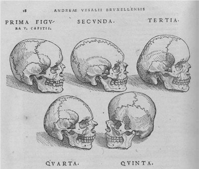
    1. Vesalius A. De Humani Corporis Fabrica. Nation Library of Medicine. :1543. http://www.nlm.nih.gov/ihm/images/A/26/919.jpg.
    1. Andrew O, M Wilkie. Craniosynostosis: genes and mechanisms. Human Molecular Genetics. 1997;6(10 Review) - PubMed
    1. Kimonis Virginia, Gold JA, Hoffman T, Panchal J, Boyadjiev S. Genetics of Craniosynostosis. Semin Pediatr Neurol. 2007;14:150–161. - PubMed

LinkOut - more resources