Review
. 2009 Jan-Mar;2(1):80-91.

The electrocardiographic profile of patients with angina pectoris

Affiliations
Review

The electrocardiographic profile of patients with angina pectoris

Carmen Ginghina et al. J Med Life. 2009 Jan-Mar.

Abstract

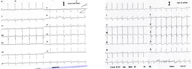
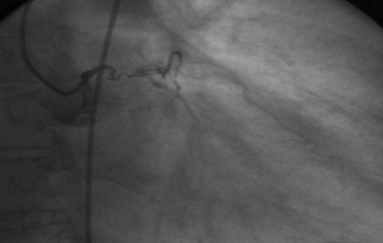
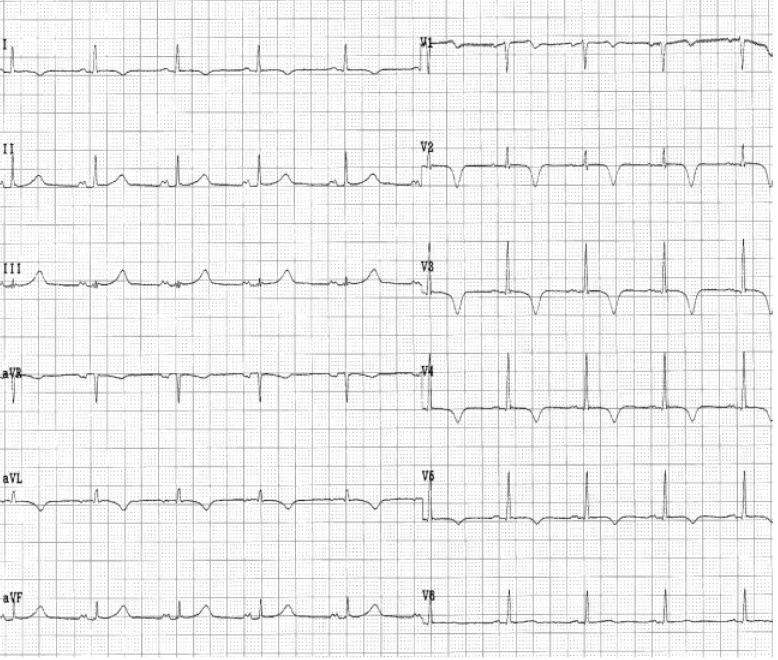
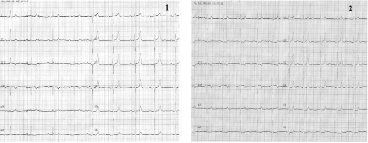
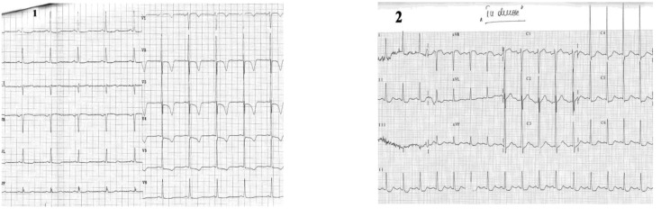
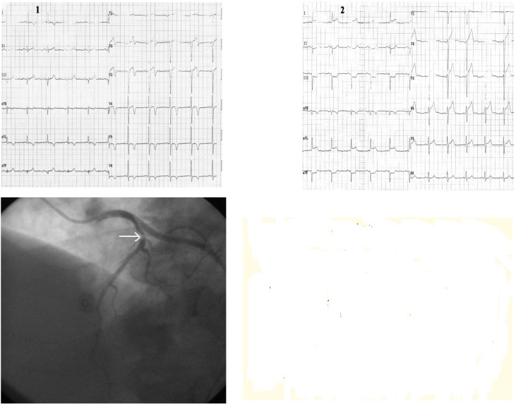
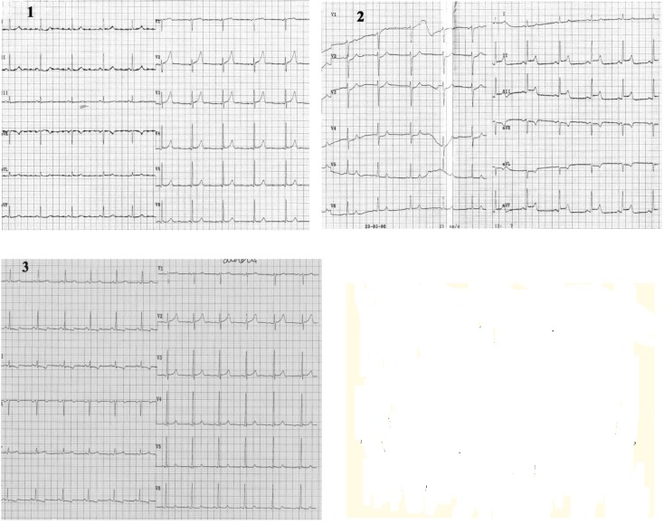
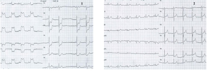
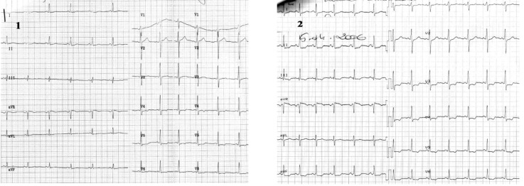
Angina pectoris is a common disabling disorder and a clinical syndrome, caused by myocardial ischemia; an imbalance between myocardial oxygen supply and myocardial oxygen consumption. Thus, ischemia produces a typical series of events such as metabolic and biochemical alterations which lead to impaired ventricular relaxation and diastolic dysfunction, impaired systolic function, and electrocardiographic abnormalities and painful symptoms of angina. Transmembrane ionic currents are responsible for the cardiac potentials that are recorded as the electrocardiogram (ECG). The electrocardiographic profile of patients with angina pectoris is variate. The electrocardiogram provides critical information for both diagnosis and prognosis, particularly when a tracing is obtained during the episodes of pain. A completely normal electrocardiogram does not exclude the possibility of acute coronary syndrome. Serial ECG tracings improve the clinician's ability to diagnose acute and chronic coronary syndromes. The ECG may assist in clarifying the differential diagnosis if taken in the presence of pain. The resting ECG also has an important role in risk stratification. Exercise ECG is more sensitive and specific than the resting ECG as far as myocardial ischemia detection is concerned, and it represents the test of choice which helps identifying inducible ischemia in the majority of patients suspected of stable angina.


Figures

Case I
Case I
Electrocardiograms of a 68 year-old woman, with prior anterior myocardial infarction and worsening of angina symptoms which became more frequent and also occurred at rest. Resting ECG (1) shows a sinus rhythm of 72 beats/min, with negative T wave in leads III and aVF. During anginal pain (2) note: ST-segment depression in leads I, aVL and V2-V6 and ST-segment elevation in lead III, aVR and V1.
None
Coronary arteriography revealed: left main chronic occlusion and a higher number of mildly stenotic and non-stenotic plaques in the right coronary artery with rich collaterally branches.
Case II
Case II
The electrocardiograms of a 62 year-old woman, corpulent, with concomitant disorders such as dyslipidemia and hypertension. She was admitted with night episodes of typical angina, with recent onset within 2 months. Resting ECG (1) shows us: a regular sinus rhythm of 70 beats/min, Q wave in leads III, aVF, negative T wave in leads III, aVF, and diphasic T wave in leads II, V2-V6, 0.5 mm ST-segment depression in leads V3-V6. During anginal pain (2) we note: increased ST - segment depression in leads V3-V6 (to 2mm) and additionally to resting ECG features, ST - segment depression in leads III and aVF. Coronary arteriography: (three vessel coronary artery disease (stenosis in the left anterior descending coronary artery (LAD) II of 90% and in LAD III of 50-60%, stenosis in the proximal intermediar coronary artery of 50% - an important vessel, and occlusion in right coronary III).
Case III
Case III
Electrocardiogram of a 62 year-old man who is admitted with new onset angina, without anterior cardiovascular diseases. The resting ECG shows: a regular sinus rhythm of 60 beats/min, AQRS +30°, negative T-wave in leads I, aVL. Echocardiographic: left ventricular antero-apical wall hypokinesia. Coronarographic: seriated stenosis in first diagonal branch of 80-90%.
Case IV
Case IV
Electrocardiograms of a 68 year-old woman, with hypertension, diabetes mellitus and dyslipidemia is hospitalized for angina pectoris provoked by rapidly increasing exertion and rest. The resting ECG (1): sinus rhythm of 60beats/min, AQRS +50°, diphasic T-wave in leads III, aVF and findings of early repolarization in precordial leads with a tall and peaked T-wave. During episode of angina pectoris 0.5 mm ST segment depression in leads II, III, aVF and V5-V6, with marked tall, peaked, positive and symmetric T-waves in precordial leads. Coronarographic: two-vessel coronary artery disease: stenosis in left anterior descending coronary artery II-III of 70-80% and in right coronary artery of 95%.
Case V
Case V
Electrocardiograms of a 72 year-old man, smoker, with hypertension, dyslipidemia, diabetes mellitus, and angina pectoris during exertion is admitted with worsening of anginal pain within the last 3 months. The ECG (1): sinus rhythm of 62 beats/min, AQRS +10°, signs of left ventricular hypertrophy, ST segment depression (max. 1.5mm) in leads V2-V6, negative T- wave in leads V1-V5. During chest pain (2) note: ST segment depression in leads I, II, aVF and V4-V6, ST segment elevation in lead aVR with positive T-waves in precordial leads. Coronarographic: three-vessel coronary artery disease: short stenosis in left anterior descending coronary artery I-II of 90%, long stenosis in left circumflex coronary artery of 90%, and occlusion in right coronary artery from origin.
Case VI
Case VI
Electrocardiograms of the 72 year-old hypertensive man admitted with angina during ordinary physical activity in the last month. The ECG at admisson (1): sinus rhythm of 63 beats/ min, AQRS -5°, negative T-wave in leads I, aVL and electrocardiographic signs of left ventricular hypertrophy with mixed abnormalities of repolarization (ST segment elevation in leads V1-V4, diphasic T-waves in leads V2-V4 and negative T-waves in leads V5-V6); During angina (2) note: ST segment elevation in leads I, aVL and V2-V5, ST segment depression in II, III, aVF, and positive T-wave in leads I, aVL, V2-V6 Coronarographic: one-vessel coronary disease: stenosis in proximal LAD of 80-90%.
Case VII
Case VII
Electrocardiograms of a 62 year-old man, smoker, with hypertension, dyslipidemia, prior coronary angioplasty with a stent in right coronary artery. He is admitted with a new onset of severe angina within the last month. The resting ECG at the admisson (1): sinus rhythm of 63beats/min, AQRS +50°, without ST-T abnormalities. During angina (2) note: ST segment elevation in leads II, III, aVF, and ST depression in leads aVL, V2-V4; at the end of chest pain (3) there are diphasic T-waves in leads II, III and aVF. Coronarographic: no significant coronary artery lesions.
Case VIII
Case VIII
Electrocardiograms of a 75 year-old man, smoker, with hypertension and stable angina pectoris for 10 years. He is admitted with angina pectoris during rest within the last 3 months. The resting ECG at the admisson (1): sinus rhythm of 68beats/min, marked (7mm) ST segment elevation in leads II, III, aVF, with positive T-wave in leads II, III, aVF, and ST segment depression in leads I, aVR, aVL, V2-V5 At the end of anginal pain note (2): decreasing in repolarization abnormalities. Coronarographic: three-vessel coronary artery disease.
Case IX
Case IX
Electrocardiograms of a 57 year-old woman, smoker, with hypertension, and on imunosupression therapy for cancer of the womb. She is admitted with 2 episodes of angina pectoris after chemotherapy procedure. The ECG, without pain (1): sinus rhythm of 92beats/min, AQRS +10°, without any repolarization abnormalities. During chest pain (2): ST segment elevation in leads I, aVL, V1-V6 and occurrence of a tall, peaked, positive T-wave in precordial leads. Coronarographic: one-vessel coronary disease: stenosis in left circumflex coronary artery of 60-70%, at the origin of the first marginal (Mg1) (arrow).
Case X
Case X
Electrocardiogram of a 68 year-old woman, with hypertension, dyslipidemia and stable angina pectoris for the last 3 years. She was admitted in hospital with typical angina with marked limitation of ordinary activities over the last month. The ECG (with pain): sinus rhythm of 130 beats/min, AQRS +45°, diffuse flat T-wave, and negative U- wave in leads V2-V6. Coronarographic: one-vessel coronary artery disease: stenosis in the proximal left anterior descending coronary artery of 75% (arrow).
Case XI
Case XI
Electrocardiograms of a 69 year-old man, with dilated cardiopathy, is admitted with chest pain and dyspnea during exertion. The ECG (patient without pain): atrial fibrillation of 100 beats/min, QS complex in leads II, III, aVF, and V2-V5 (1). During pain: ST segment depression in leads I, aVL, V2-V6, prolonged QTc interval (QTc = 520msec) and diphasic T-wave in leads V2-V6 (2). Coronarographic: three-vessel coronary artery disease (stenosis in LADII of 80%, stenosis in Mg 1 of 70% and occlusion in right coronary artery).
Case XII
Case XII
Electrocardiograms of a 75 years old man, corpulent, with dyslipidemia, hypertension, diabetes mellitus, prior myocardial infarction, is admitted with angina pectoris at rest. The ECG without pain (1): sinus rhythm of 93 beats/min, AQRS +10°, ST segment depression in leads I, aVL, V3-V6. During pain (2): increased ST segment depression and occurrence of negative T-wave in I, aVL, V4-V6, and ST segment elevation in III, aVR. Coronarographic: three-vessel coronary disease (stenosis in LAD II of 90%, occlusion in Cx, hypoplasia of right coronary artery.
None
Electrocardiograms of a 38 year-old female, without a previous cardiac disease, is admitted with severe chest pain new onset within the last 3 weeks. The ECG without pain (1): sinus rhythm of 92 beats/min, AQRS +30°, minor ST- segment depression in leads I, II, V4-V6. During chest pain (2): ST segment depression in leads III, aVF, V3 , increased ST-segment depression in leads II, V4-V6, with ST-segment elevation in aVR . Coronarographic: no significant coronary artery lesions.

Similar articles

Cited by

References

    1. C Ginghina, M Marinescu, D Dragomir. Indreptar de diagnostic si tratament in infarctul miocardic acut. 2002:77–123.
    1. Zipes , Libby , Bonow , Braunwald Coronary blood flow and myocardial iaschemia. 2005;44:1103–1107.
    1. Gibbons RJ, Abrams J, Chatterjee K, et al. ACC/AHA 2002 guideline update for the management of patients with chronic stable angina : a report of the American College of Cardiology /American Heart Association Task Force on Practice Guidelines ( Committee to Update the 1999 Guidelines for the Management of Patient with Chronic Stable Angina ) J Am Coll Cardiol. 2003 Jan 1;41(1):159–168. - PubMed
    1. Kim Fox, et al. Guidelines on the management of stable angina pectoris : the task force on the management of stable angina pectoris of the European Society of Cardiology. Eur Heart J. 2006;10:10–93. - PubMed
    1. Diderholm E, Andren B, et al. ST depression in ECG at entry indicates severe coronary lesions and large benefits of an early invasive treatment strategy in unstable coronary artery disease; the FRISC II ECG substudy. The Fast Revascularisation during InStability in Coronary artery disease. Eur Heart J. 2002 Jan;23(1):41–49. - PubMed

LinkOut - more resources