

The development of xenograft glioblastoma implants in nude mice brain
- PMID: 20108505
- PMCID: PMC3018968
The development of xenograft glioblastoma implants in nude mice brain
Abstract
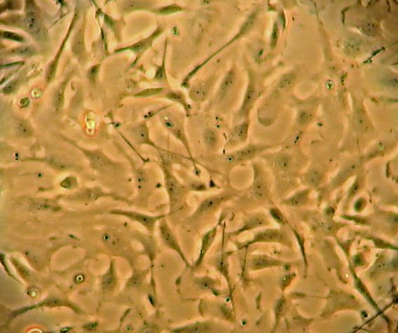
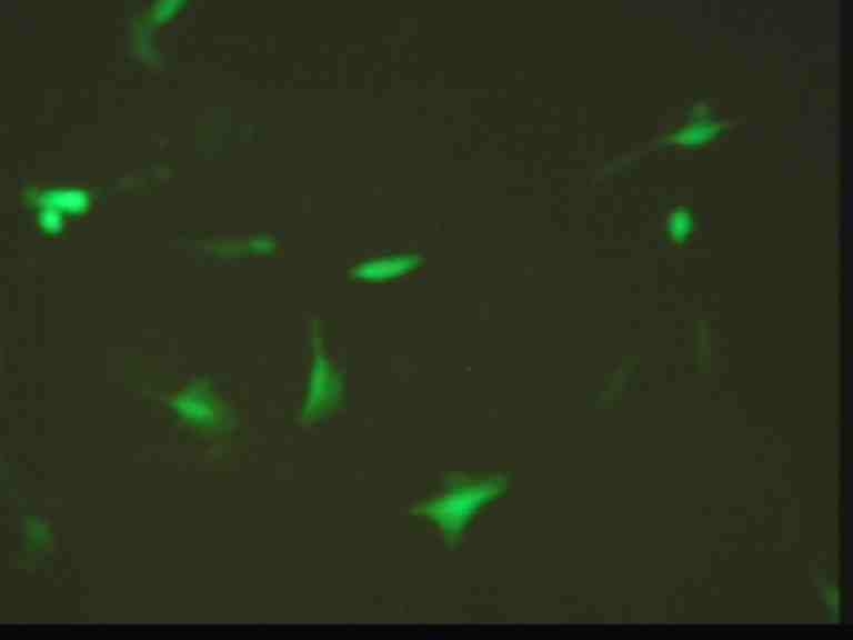
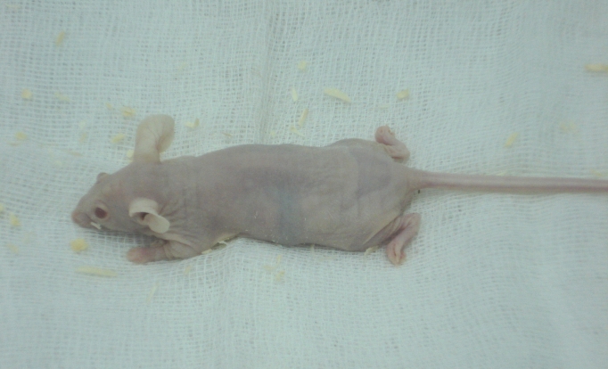
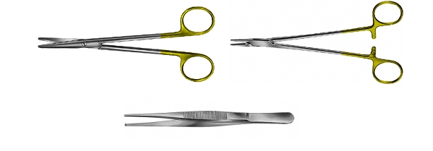
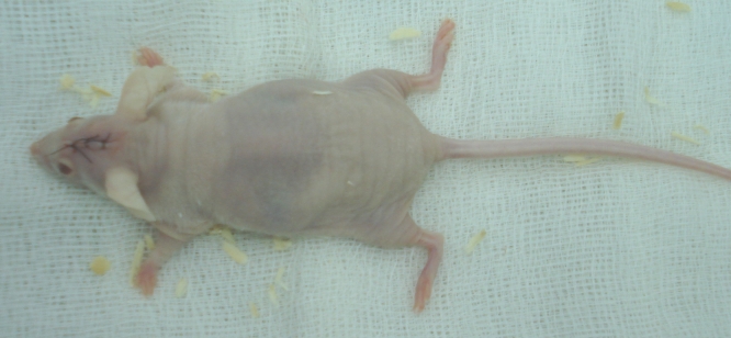
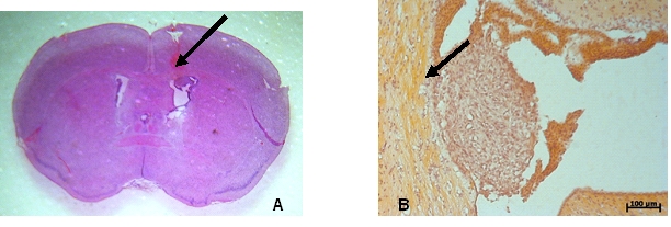
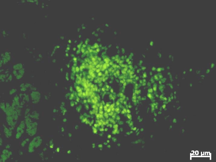
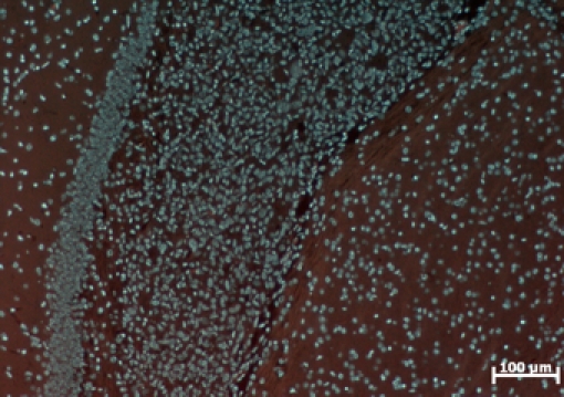
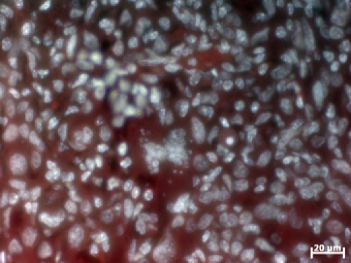
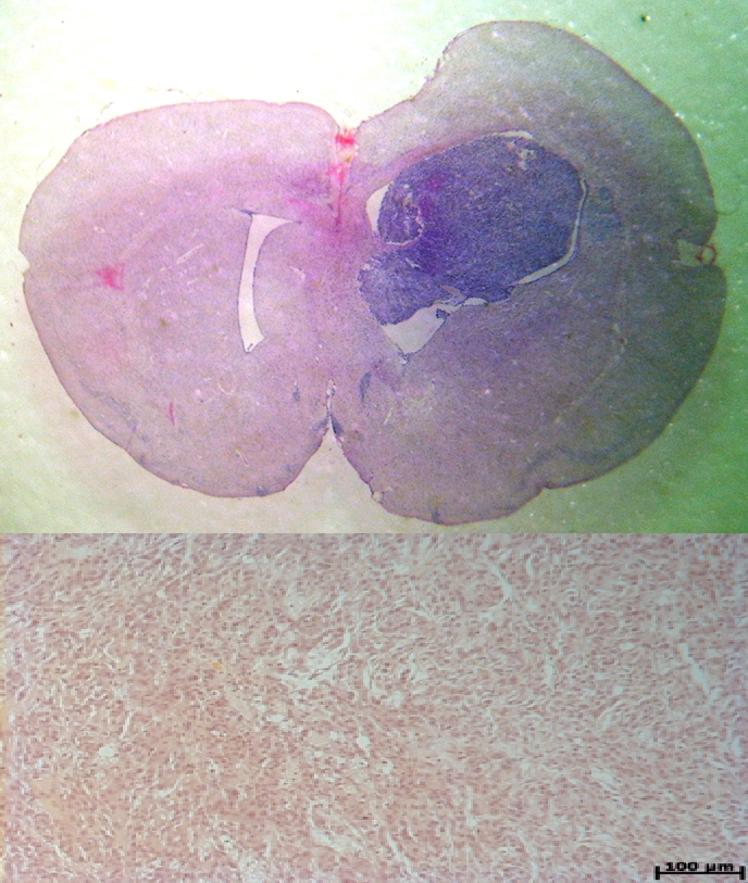
The inefficacity of the actual therapies for glioblastoma multiformis stimulates the researchers to search for new and innovative therapies. Therefore, the development of in vivo model for glioblastoma is an essential step during these researches, being a link between cells cultures studies and the first phases of clinical trials. In this paper, we present several procedures which have been performed for the first time in our country, such as: the cultivation and manipulation of U87MG line, the manipulation of athymic knock-out mice (NUDE Crl: CD-1 Foxn 1), the stereotactic inoculation of glioblastoma cells and finally the development of glioblastoma xenograft in the brain of inoculated nude mice. These results, which offer to the researchers from our country an in vivo model for glioblastoma, could be the start point for several projects oriented to the development of new therapies in glioblastoma, and could raise the performance of our scientific research to the European level.
Figures
Similar articles
-
Stereotactic intracranial implantation and in vivo bioluminescent imaging of tumor xenografts in a mouse model system of glioblastoma multiforme.J Vis Exp. 2012 Sep 25;(67):4089. doi: 10.3791/4089. J Vis Exp. 2012. PMID: 23051742 Free PMC article.
-
Xenograft transplantation of human malignant astrocytoma cells into immunodeficient rats: an experimental model of glioblastoma.Clinics (Sao Paulo). 2010 Mar;65(3):305-9. doi: 10.1590/S1807-59322010000300011. Clinics (Sao Paulo). 2010. PMID: 20360922 Free PMC article.
-
The growth of glioblastoma orthotopic xenografts in nude mice is directly correlated with impaired object recognition memory.Physiol Behav. 2014 Jan 17;123:55-61. doi: 10.1016/j.physbeh.2013.09.012. Epub 2013 Oct 2. Physiol Behav. 2014. PMID: 24096193
-
[Heterotransplantation of a human glioma and brain metastases in the athymic nude mouse--a preclinical model for radiation oncology. 1. Basic principles and methodology].Strahlenther Onkol. 1988 Apr;164(4):235-43. Strahlenther Onkol. 1988. PMID: 2834833 Review. German.
-
Metastasis along the stereotactic biopsy trajectory in glioblastoma multiforme.Acta Neurochir (Wien). 1999;141(9):1011-2. doi: 10.1007/s007010050410. Acta Neurochir (Wien). 1999. PMID: 10526085 Review. No abstract available.
Cited by
-
The orthotopic xenotransplant of human glioblastoma successfully recapitulates glioblastoma-microenvironment interactions in a non-immunosuppressed mouse model.BMC Cancer. 2014 Dec 8;14:923. doi: 10.1186/1471-2407-14-923. BMC Cancer. 2014. PMID: 25482099 Free PMC article.
-
Development of a Prodrug of Camptothecin for Enhanced Treatment of Glioblastoma Multiforme.Mol Pharm. 2021 Apr 5;18(4):1558-1572. doi: 10.1021/acs.molpharmaceut.0c00968. Epub 2021 Mar 1. Mol Pharm. 2021. PMID: 33645231 Free PMC article.
-
Spectral tracing of deuterium for imaging glucose metabolism.Nat Biomed Eng. 2019 May;3(5):402-413. doi: 10.1038/s41551-019-0393-4. Epub 2019 Apr 29. Nat Biomed Eng. 2019. PMID: 31036888 Free PMC article.
-
Highly Invasive Fluorescent/Bioluminescent Patient-Derived Orthotopic Model of Glioblastoma in Mice.Front Oncol. 2022 Jul 13;12:897839. doi: 10.3389/fonc.2022.897839. eCollection 2022. Front Oncol. 2022. PMID: 35912166 Free PMC article.
-
Preclinical assessment of thrombin-preconditioned human Wharton's jelly-derived mesenchymal stem cells for neonatal hypoxic-ischaemic brain injury.J Cell Mol Med. 2021 Nov;25(22):10430-10440. doi: 10.1111/jcmm.16971. Epub 2021 Oct 15. J Cell Mol Med. 2021. PMID: 34651412 Free PMC article.
References
-
- Brehar FM, Ciurea AV, Tascu A, Gherghina E, Ciurel I. Isolation and characterization of a human glioblastoma cell line. Romanian Neurosurgery. 2007
-
- Andreansky S, Soroceanu L, Flotte ER. Evaluation of genetically engineered herpes simplex viruses as oncolytic agents for human malignant brain tumors. Cancer Res . 1997;57:1502–1509. - PubMed
-
- Friedman HS, Dolan ME, Pegg AE. Activity of temozolomide in the treatment of central nervous system tumor xenografts. Cancer Res. 1995;55:2853–2857. - PubMed
-
- Friedman HS, Keir ST, Houghton PJ. Activity of irofulven (6 –hydroxymethylacylfulvene) in the treatment of glioblastoma multiforme–derived xenografts in athymic mice. Cancer Chemother Pharmacol . 2001;48:413–416. - PubMed
-
- Kaye AH, Morstyn G, Gardner I. Development of a xenograft glioma model in mouse brain. Cancer Res . 1986;46:1367–1373. - PubMed
Publication types
MeSH terms
LinkOut - more resources
Full Text Sources
Medical