

The management of primitive retroperitoneal tumors--problems of clinical, imaging diagnosis, and treatment
- PMID: 20108510
- PMCID: PMC5654303
The management of primitive retroperitoneal tumors--problems of clinical, imaging diagnosis, and treatment
Abstract
Retroperitoneal tumors, whether primary or resulting from the metastasis of other tumors, are a real challenge for the surgeon, in terms of their diagnosis and treatment. They are relatively rare, under 0.2% of the total number of tumors. The clinical examination of retroperitoneal tumors is uncharacteristic and misleading, consisting mainly in palpation of the tumor proper and in assessment of pain. The other signs and symptoms often result from the affected neighboring organs. The imaging investigations used in diagnosing retroperitoneal tumors are ecography, intravenous pyelography, computed tomography, MRI, PET/CT. The main treatment is surgical, consisting either in total or partial excision of the tumor, or in biopsy samples to make a histopathologic diagnosis. Post-operative course depends mainly on the thoroughness of the surgical treatment, that is the complete excision of the tumor, which increases the chances of survival, while lowering the risk of relapse.
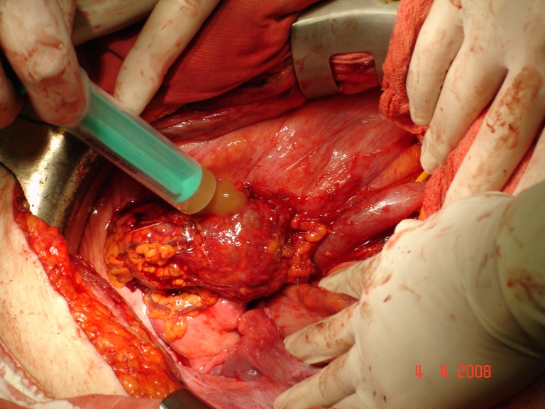
Figures


References
-
- Setlacec D., Proca E., Popa C. Primitive Retroperitoneal Tumors. Bucuresti: Ed. Medicala; 1986 .
-
- Sinescu I., Gluck G., Harza M. Oncologic Urology, vol. 2. Bucuresti: Ed. Universitara “Carol Davila”; 2006 . pp. 627–645.
-
- Manu M, Neicutescu C, Hortopan M, Proca E, Sinescu I. Clinical, Imagistic and Histopathologic Study and Therapeutic Approach in Retroperitoneal Tumors. Revista Romana de Urologie. 2005;4(1):5–8.
-
- Mischianu D, Bana M, Dinu M, Vlasin G. Primitive Retroperitoneal Tumors –from the Experience of the Urology Clinic of the Central Military Hospital. Revista Chirurgia, Ed. Celsius, Bucuresti. 2002;97(2):139–150. - PubMed
-
- Lucan M. Treaty of Surgery, Diseases of the Retroperitoneum, edited by Irinel Popescu, vol. I “Urologie”. Bucuresti: Ed. Academiei Romane; 2007. pp. 71–77.
Publication types
MeSH terms
LinkOut - more resources
Full Text Sources