. 2010 Mar 9;49(9):1822-32.
doi: 10.1021/bi901974a.

Mechanism and specificity of a symmetrical benzimidazolephenylcarboxamide helicase inhibitor

Affiliations

Mechanism and specificity of a symmetrical benzimidazolephenylcarboxamide helicase inhibitor

Craig A Belon et al. Biochemistry. .

Abstract

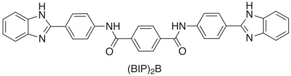
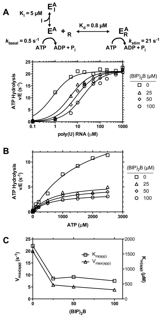
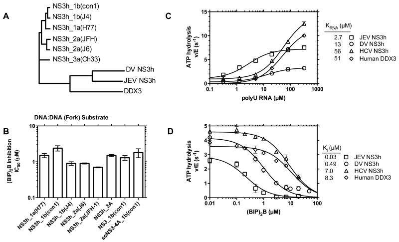
This study examines the effects of 1-N,4-N-bis[4-(1H-benzimidazol-2-yl)phenyl]benzene-1,4-dicarboxamide ((BIP)(2)B) on the NS3 helicase encoded by the hepatitis C virus (HCV). Molecular beacon-based helicase assays were used to show that (BIP)(2)B inhibits the ability of HCV helicase to separate a variety of RNA and DNA duplexes with half-maximal inhibitory concentrations ranging from 0.7 to 5 microM, depending on the nature of the substrate. In single turnover assays, (BIP)(2)B only inhibited unwinding reactions when it was preincubated with the helicase-nucleic acid complex. (BIP)(2)B quenched NS3 intrinsic protein fluorescence with an apparent dissociation constant of 5 microM, and in the presence of (BIP)(2)B, HCV helicase did not appear to interact with a fluorescent DNA oligonucleotide. In assays monitoring HCV helicase-catalyzed ATP hydrolysis, (BIP)(2)B only inhibited helicase-catalyzed ATP hydrolysis in the presence of intermediate concentrations of RNA, suggesting RNA and (BIP)(2)B compete for the same binding site. HCV helicases isolated from various HCV genotypes were similarly sensitive to (BIP)(2)B, with half-maximal inhibitory concentrations ranging from 0.7 to 2.4 microM. (BIP)(2)B also inhibited ATP hydrolysis catalyzed by related helicases from Dengue virus, Japanese encephalitis virus, and humans. (BIP)(2)B appeared to bind the HCV and human proteins with similar affinity (K(i) = 7 and 8 microM, respectively), but it bound the flavivirus proteins up to 270 times more tightly. Results are discussed in light of a molecular model of a (BIP)(2)B-HCV helicase complex, which is unable to bind nucleic acid, thus preventing the enzyme from separating double-stranded nucleic acid.


Figures

Figure 1
Figure 1
1-N,4-N-bis[4-(1H-benzimidazol-2-yl)phenyl]benzene-1,4-dicarboxamide, (BIP)2B.
Figure 2
Figure 2
Effect of (BIP)2B on HCV helicase catalyzed DNA and RNA unwinding. (A) MBHA substrates used in this study. Regions forming hairpin structures after strand separation are underlined. (B) A representative MBHA using the DNA:DNA substrate in the presence (circles) or absence (squares) of 25 μM (BIP)2B. The arrow shows when ATP was added. Error bars represent standard deviations of three independent reactions. (C) Inhibition of HCV NS3h catalyzed substrate unwinding by (BIP)2B. Fluorescence measurements at time zero, F0, and 30 minutes, F30, are used to compute Inhibition% with Equation 1, which is plotted for the DNA:DNA (fork) substrate versus (BIP)2B concentration. Error bars represent the standard deviation of three independent reactions. Points are fit to Equation 2. (D) Concentration of (BIP)2B required to inhibit 50 % of NS3h catalyzed strand separation in MBHAs using indicated substrates. IC50 values plotted are averages of three values that were determined from three independent titrations with (BIP)2B. Errors are standard deviations.
Figure 3
Figure 3
Effect of (BIP)2B on HCV catalyzed DNA unwinding in the presence of a protein trap (single turnover conditions). Pre-steady state time courses for NS3h_1b (con1) catalyzed DNA:DNA MBHAs performed in the presence of 0, 0.5, 2.5 or 10 μM (BIP)2B and 1 μM dT20 (enzyme trap). All time courses shown represent the average of four independent experiments. Error bars are omitted for clarity. (A) A syringe containing enzyme, DNA and (BIP)2B is mixed with a syringe containing ATP and dT20 to initiate unwinding such that final concentrations of all reagents are the same as in a standard helicase reaction. (B) A syringe containing enzyme and DNA (preformed E-DNA complex) is mixed with a syringe containing (BIP)2B, ATP and dT20 to initiate unwinding. Data are fit to equation 3 with the constants in Table I.
Figure 4
Figure 4
Interaction of (BIP)2B with enzyme in the absence or presence of DNA. (A) Effect of (BIP)2B on NS3h protein fluorescence. The protein fluorescence of a constant concentration of enzyme is measured as a solution is titrated with (BIP)2B. Corrected fluorescence (F, Equation 4) of three different NS3h_1b (con1) solutions was globally fit to Equation 5, which assumes a different fluorescence for free enzyme and an enzyme-inhibitor complex described by the coefficients Ff and Fc, respectively. In (B), the fluorescence of 2 nM F18 DNA (5′-GCC TCG CTG CCG TCG CCA-FAM-3′) was measured while titrating with HCV NS3h_1b (con1) in the presence of various concentrations of (BIP)2B. Data were globally fit to Equation 6 to provide an estimate of Ki for the NS3h-(BIP)2B complex formation and a Kd for the NS3h-DNA complex.
Figure 5
Figure 5
Effects of (BIP)2B on helicase catalyzed ATP hydrolysis. (A) Stimulation of NS3h_1b (con1) catalyzed ATP hydrolysis by poly(U) RNA in the presence of various concentrations of (BIP)2B. Note that basal ATPase rate (in the absence of RNA) and maximum ATPase rates are not affected by (BIP)2B. Only at intermediate concentrations of RNA does (BIP)2B inhibit ATPase activity. Data are globally fit between data sets using Equation 7 to estimate the constants shown. (B) Effect of (BIP)2B on ATP hydrolysis at various ATP concentrations. Reactions were performed with NS3h_1b (con1) at various ATP concentrations in the presence of 15 μM poly(U). Data are fit between to Equation 8. (C) Subplot of apparent Km's and Vmax's determined from fits in panel B.
Figure 6
Figure 6
Effects of (BIP)2B on helicases encoded by various HCV genotypes, flaviviruses and humans. (A) Phylogenetic tree of the hepatitis C virus NS3h proteins used in this study along with NS3h from dengue virus (DV), NS3h from Japanese encephalitis virus (JEV) and human DDX3 helicase. The tree was drawn from amino acid alignments generated with CLC Sequence Viewer (http://www.clcbio.com). (B) Inhibition of HCV helicase catalyzed DNA:DNA(fork) unwinding by (BIP)2B, reported as IC50 values with error bars of standard deviation of three independent determinations. “NS3h_x(y)” indicates a truncated NS3 protein lacking a protease domain of HCV genotype x (isolate y). NS3_1b(con1) is the full length NS3 protein and scNS3-4A_1b(con1) is a full length NS3 protein with a covalently linked HCV NS4A peptide. (C) RNA-stimulated ATP hydrolysis catalyzed by HCV, JEV, DV and DDX3 helicases at pH 7.5. The viral HCV, JEV and DV helicases all exhibit non-zero basal ATPase rates in the absence of RNA while human DDX3 helicase does not detectably hydrolyze ATP in the absence of RNA. Data are fit to Equation 9 with each point representing the average of three independent experiments; error bars show standard deviations. (D) Inhibition of ATP hydrolysis by (BIP)2B when HCV, JEV, DV and DDX3 helicases are partially stimulated by 15 μM RNA. Data are fit to an equation describing competitive inhibition (Equation 10) with each point representing the average of three independent experiments; error bars show standard deviations.
Figure 7
Figure 7
Molecular model of (BIP)2B bound to the HCV NS3h protein. UCSF DOCK 6.3 was used to bind (BIP)2B to the oligonucleotide binding site of PDB structure 1A1V (9). NS3h domains 1, 2 and 3 are represented by yellow, magenta and tan ribbons, respectively. Bound ligands from PDB 1A1V are shown as red sticks (oligonucleotide and a bound sulfate), and the docked (BIP)2B molecule is shown as a space filling model colored by atom. The insert shows a close-up view of the putative (BIP)2B binding site with nearby amino acid side chains labeled.

Similar articles

Cited by

References

    1. Kwong AD, Rao BG, Jeang KT. Viral and cellular RNA helicases as antiviral targets. Nat Rev Drug Discov. 2005;4:845–853. - PMC - PubMed
    1. Frick DN, Lam AM. Understanding helicases as a means of virus control. Curr Pharm Des. 2006;12:1315–1338. - PMC - PubMed
    1. Frick DN. The hepatitis C virus NS3 protein: a model RNA helicase and potential drug target. Curr Issues Mol Biol. 2007;9:1–20. - PMC - PubMed
    1. Belon CA, Frick DN. Helicase inhibitors as specifically targeted antiviral therapy for hepatitis C. Future Virol. 2009;4:277–293. - PMC - PubMed
    1. Phoon CW, Ng PY, Ting AE, Yeo SL, Sim MM. Biological evaluation of hepatitis C virus helicase inhibitors. Bioorg Med Chem Lett. 2001;11:1647–1650. - PubMed

Publication types

MeSH terms

Substances