. 2010 Jan 28:11:10.
doi: 10.1186/1471-2121-11-10.

Mechano-transduction in periodontal ligament cells identifies activated states of MAP-kinases p42/44 and p38-stress kinase as a mechanism for MMP-13 expression

Affiliations

Mechano-transduction in periodontal ligament cells identifies activated states of MAP-kinases p42/44 and p38-stress kinase as a mechanism for MMP-13 expression

Nelli Ziegler et al. BMC Cell Biol. .

Abstract

Background: Mechano-transduction in periodontal ligament (PDL) cells is crucial for physiological and orthodontic tooth movement-associated periodontal remodelling. On the mechanistic level, molecules involved in this mechano-transduction process in PDL cells are not yet completely elucidated.

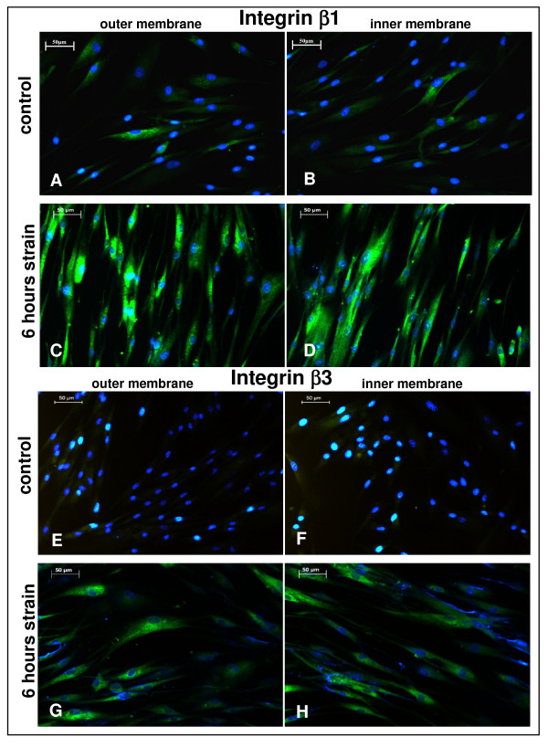
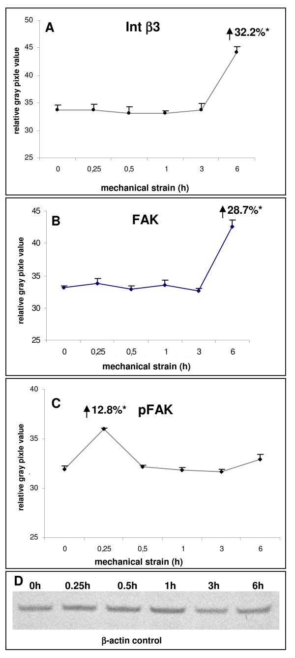
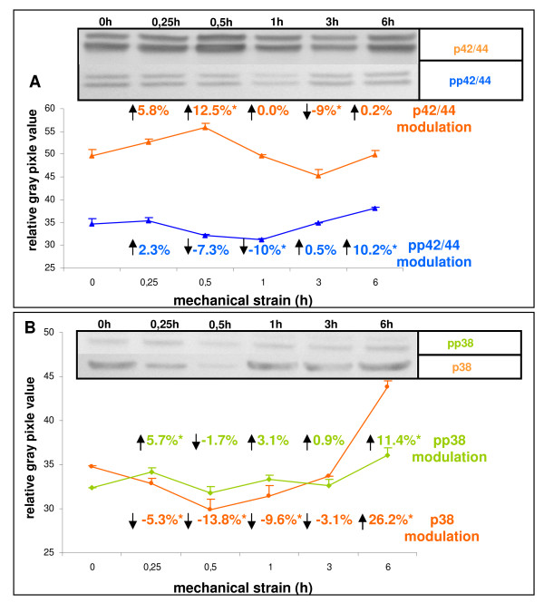
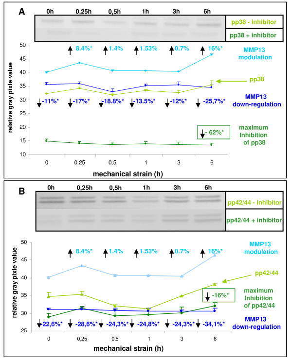
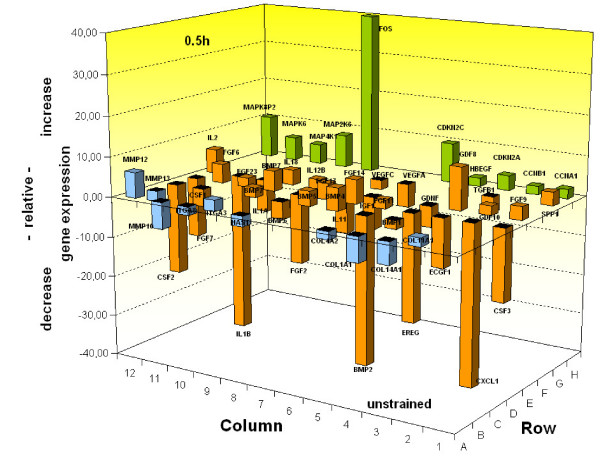
Results: In the present study we show by western blot (WB) analysis and/or indirect immunofluorescence (IIF) that mechanical strain modulates the amount of the matrix metalloproteinase MMP-13, and induces non-coherent modulation in the amount and activity of signal transducing molecules, such as FAK, MAP-kinases p42/44, and p38 stress kinase, suggesting their mechanistic role in mechano-transduction. Increase in the amount of FAK occurs concomitant with increased levels of the focal contact integrin subunits beta3 and beta1, as indicated by WB or optionally by IIF. By employing specific inhibitors, we further identified p42/44 and p38 in their activated, i.e. phosphorylated state responsible for the expression of MMP-13. This finding may point to the obedience in the expression of this MMP as extracellular matrix (ECM) remodelling executioner from the activation state of mechano-transducing molecules. mRNA analysis by pathway-specific RT-profiler arrays revealed up- and/or down-regulation of genes assigning to MAP-kinase signalling and cell cycle, ECM and integrins and growth factors. Up-regulated genes include for example focal contact integrin subunit alpha3, MMP-12, MAP-kinases and associated kinases, and the transcription factor c-fos, the latter as constituent of the AP1-complex addressing the MMP-13 promotor. Among others, genes down-regulated are those of COL-1 and COL-14, suggesting that strain-dependent mechano-transduction may transiently perturbate ECM homeostasis.

Conclusions: Strain-dependent mechano-/signal-transduction in PDL cells involves abundance and activity of FAK, MAP-kinases p42/44, and p38 stress kinase in conjunction with the amount of MMP-13, and integrin subunits beta1 and beta3. Identifying the activated state of p42/44 and p38 as critical for MMP-13 expression may indicate the mechanistic contribution of mechano-transducing molecules on executioners of ECM homeostasis.


Figures

Figure 1
Figure 1
Strain-induced integrin β1 and β3 expression in periodontal ligament (PDL) cells after 6 hours. (A) PDL cells were seeded on flexible bottom cell culture dishes and strained with averaged 2.5% for 6 hours. For indirect immunofluorescence (IIF), the flexible membrane was divided into pieces derived from the inner and outer part of the membrane, and fixed for staining procedure. In IIF integrin subunit β1 (A, B) and subunit β3 (E, F) were visualized in controls and after 6 hours of strain-application (β1 C, D and β3 G, H) by using an anti-integrin β1 and β3-specific antibody. The green fluorescent signal illustrates the integrin β1 and β3 expression, while the blue fluorescence visualises the nuclear DAPI-staining. Scale bar, 50 μm.
Figure 2
Figure 2
Immunoblotting analysis of mechano-sensitive molecular constituents of the focal adhesion complex in PDL cells after strain-application. PDL cells were seeded on flexible bottom cell culture dishes, strained with averaged 2.5% and followed by westernblot analysis. The numerical expression values were always denoted in relation to unstrained controls. (A) Expression levels of integrin β3 in PDL cells in response of the strain kinetic were detected with a mouse monoclonal anti-integrin β3 antibody and the maximum expression modulation was numerical notified in the graph after 6 h of strain. (B) For detection of the strain-induced total protein expression level of the focal adhesion kinase (FAK), a polyclonal rabbit anti-FAK antibody was used, and the maximum expression value was marked in the graph after 6 h strain. (C) Phosphorylation levels of Tyr576 of FAK were assessed by immunoblotting using an antibody against phospho-Tyr576. The maximum activation status was numerically denoted after 0.25 h strain. (D) In addition, for loading control and for internal normalisation, β-actin was detected for each immunoblotting experiment. Data of each graph represent the mean of three individual experiments (n = 3), mean +/- SD and means were subjected to the Students T-test. All compared mean values with p < 0.01 were considered as statistical significant and are marked with an asterisk.
Figure 3
Figure 3
Immunoblotting of molecules involved in mechano-signal-transduction. PDL cells were seeded on flexible bottom cell culture dishes, strained with averaged 2.5% and followed by westernblot analysis. The numerical expression values were always denoted in relation to unstrained controls (A) The modulation of expression levels of the MAP-kinases p42/44 following strain application were detected with the monoclonal rabbit anti-p42/44 antibody, and the numerical changes of increase and decrease in expression were denoted by arrows in the graph. The phosphorylation levels of Thr202/Tyr204 of p42/44 were assessed by immunoblotting, using an antibody against phospho- Thr202/Tyr204. The numerical changes in the activation status of pp42/44 in consequence of strain application were visualised by arrows in the graph. (B) The strain-induced expression levels of the MAP-kinase p38 were detected with a monoclonal rabbit anti-p38 antibody, and the expression changes increase or decrease were marked in the graph by arrows. The strain-induced activation status of phospho-p38 (pp38) was detected by immunoblotting using an antibody against phospho- Thr180/Tyr182 of p38, and the modulation of expression levels was notified with numerical values and arrows in the graph. Data of each graph represent the mean of three individual experiments (n = 3), mean +/- SD and means were subjected to the Students T-test. All compared mean values with p < 0.01 were considered as statistical significant and are marked with an asterisk. The depicted western blots exemplify the protein expression changes of one biological replicate.
Figure 4
Figure 4
Role of activated MAP-kinases pp38 and pp42/44 in strain-induced expression of the active form of MMP-13. PDL cells were seeded on flexible bottom cell culture dishes, strained with averaged 2.5% and followed by westernblot analysis. The numerical expression values were denoted in relation to unstrained controls. The maximum inhibition was related to the respective strain-kinetic time points. (A) For strain-induced MMP-13 expression detection in a pp38-specific inhibition assay, PDL cells were pre-treated with 10 μM of a phosphorylation-specific inhibitor (SB202190) against pp38 and the MMP-13 down-regulation was compared versus untreated control. The numerical changes of the MMP13 protein expression status in consequence of strain application were visualised by arrows in the graph. Efficiency of pp38 inhibition yielded in a maximum inhibition of 62% after 6 hours strain and was visualised by the immunoblots (inset). (B) Strain-induced expression of MMP-13 in a pp42/44-specific inhibition assay was performed by pre-treating PDL cells with 10 μM of a phosphorylation-specific inhibitor (UO-126) against pp42/44 and the MMP-13 down-regulation was compared versus untreated control. The numerical changes of the MMP-13 protein expression status in consequence of strain application were visualised by arrows in the graph. Efficiency of pp42/44 inhibition yielded in a maximum inhibition of 16% after 6 hours strain and was visualised by the immunoblots (inset). Data of each graph represent the mean of three individual experiments (n = 3), mean +/- SD and means were subjected to the Students T-test. All compared mean values with p < 0.01 were considered as statistical significant and are marked with an asterisk. The depicted western blots exemplify the protein expression changes of one biological replicate.
Figure 5
Figure 5
Strain-induced quantitative mRNA-expression analysis of PDL cells related to MAP-kinase signalling and cell cycle, ECM and integrins, and growth factor pathways. PDL cells were seeded on flexible bottom cell culture dishes and strained with averaged 2.5%. For quantitative mRNA analysis, PDL cells of unstrained controls and cells after 0.5 h strain period were harvested, and RNA was isolated and quantified for pathway-specific analysis. The relative increase and decrease in gene expression were internally normalised versus a quotient of 4 different housekeeping genes, and plotted in the graph. The data represent the mean of three independent experiments, and only significant expression modulations were included in the graph. The respective statistics were considered in the evaluation software. Plotted genes were allocated to a specific pathway and the respective columns were coloured for MAP-kinase signalling and cell cycle in orange, for ECM and integrins in light blue, and for growth factors in green, respectively.

Similar articles

Cited by

References

    1. von Bohl M, Kuijpers-Jagtman AM. Hyalinization during orthodontic tooth movement: a systematic review on tissue reactions. Eur J Orthod. 2009;31:30–36. doi: 10.1093/ejo/cjn080. - DOI - PubMed
    1. Bartold PM, McCulloch CA, Narayanan AS, Pitaru S. Tissue engineering: a new paradigm for periodontal regeneration based on molecular and cell biology. Periodontol. 2000;24:253–269. doi: 10.1034/j.1600-0757.2000.2240113.x. - DOI - PubMed
    1. Cho MI, Garant PR. Development and general structure of the periodontium. Periodontol. 2000;24:9–27. doi: 10.1034/j.1600-0757.2000.2240102.x. - DOI - PubMed
    1. Tsuruga E, Sato A, Ueki T, Nakashima K, Nakatomi Y, Ishikawa H, Yajima T, Sawa Y. Integrin alphavbeta3 regulates microfibril assembly in human periodontal ligament cells. Tissue Cell. 2009;41:85–89. doi: 10.1016/j.tice.2008.07.005. - DOI - PubMed
    1. Himmel M, Ritter A, Rothemund S, Pauling BV, Rottner K, Gingras AR, Ziegler WH. Control of high affinity interactions in the talin C-terminus - how talin domains coordinate protein dynamics in cell adhesions. J Biol Chem. 2009;284:13832–13842. doi: 10.1074/jbc.M900266200. - DOI - PMC - PubMed

Publication types

MeSH terms

LinkOut - more resources