. 2010 Jan 28:7:7.
doi: 10.1186/1742-2094-7-7.

Acute paretic syndrome in juvenile White Leghorn chickens resembles late stages of acute inflammatory demyelinating polyneuropathies in humans

Affiliations

Acute paretic syndrome in juvenile White Leghorn chickens resembles late stages of acute inflammatory demyelinating polyneuropathies in humans

Sophie R Bader et al. J Neuroinflammation. .

Abstract

Background: Sudden limb paresis is a common problem in White Leghorn flocks, affecting about 1% of the chicken population before achievement of sexual maturity. Previously, a similar clinical syndrome has been reported as being caused by inflammatory demyelination of peripheral nerve fibres. Here, we investigated in detail the immunopathology of this paretic syndrome and its possible resemblance to human neuropathies.

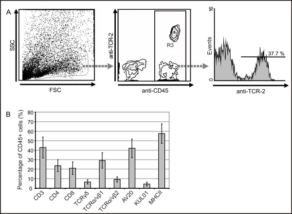
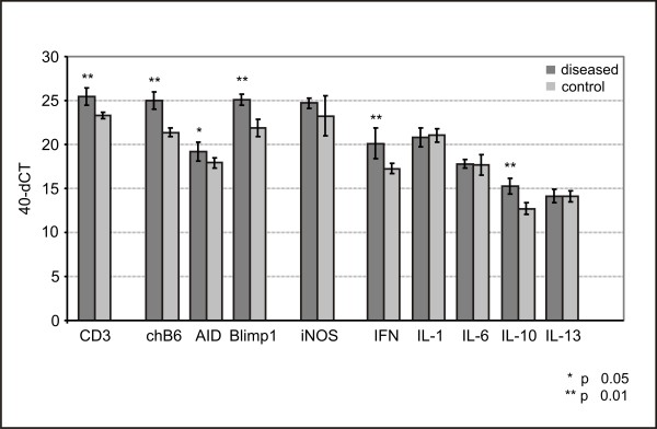
Methods: Neurologically affected chickens and control animals from one single flock underwent clinical and neuropathological examination. Peripheral nervous system (PNS) alterations were characterised using standard morphological techniques, including nerve fibre teasing and transmission electron microscopy. Infiltrating cells were phenotyped immunohistologically and quantified by flow cytometry. The cytokine expression pattern was assessed by quantitative real-time PCR (qRT-PCR). These investigations were accomplished by MHC genotyping and a PCR screen for Marek's disease virus (MDV).

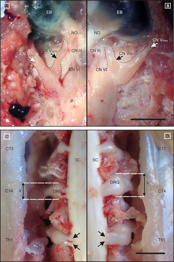
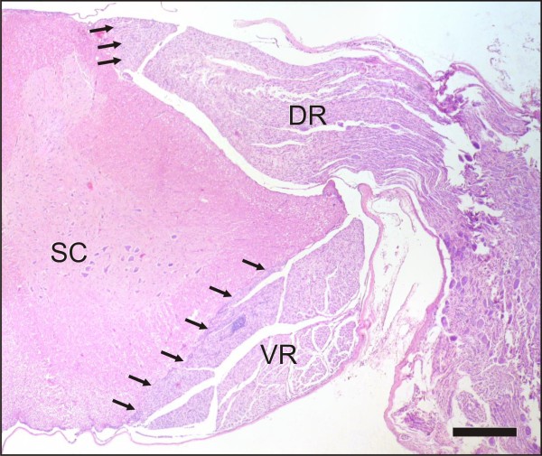
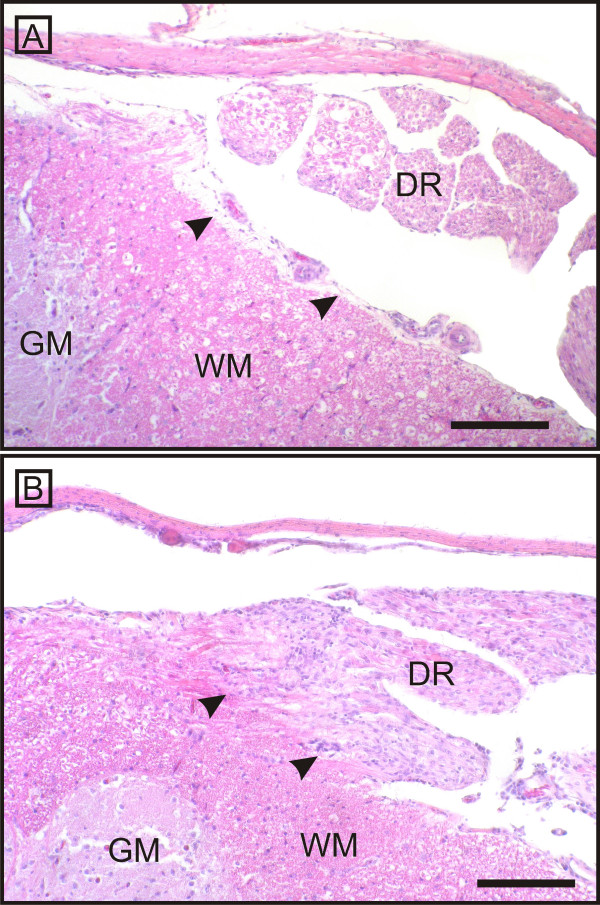
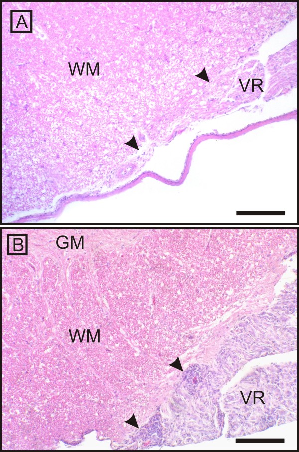
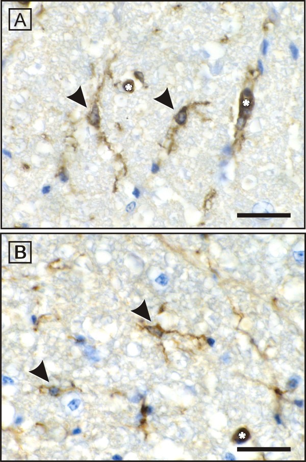
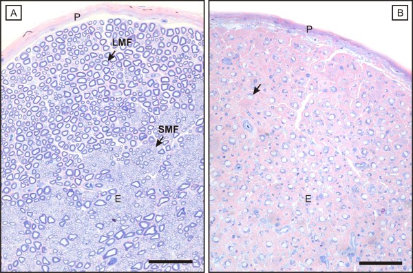
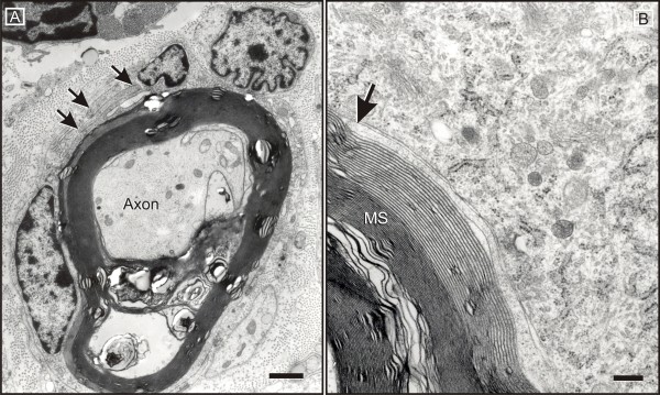
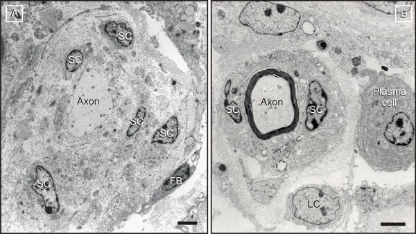
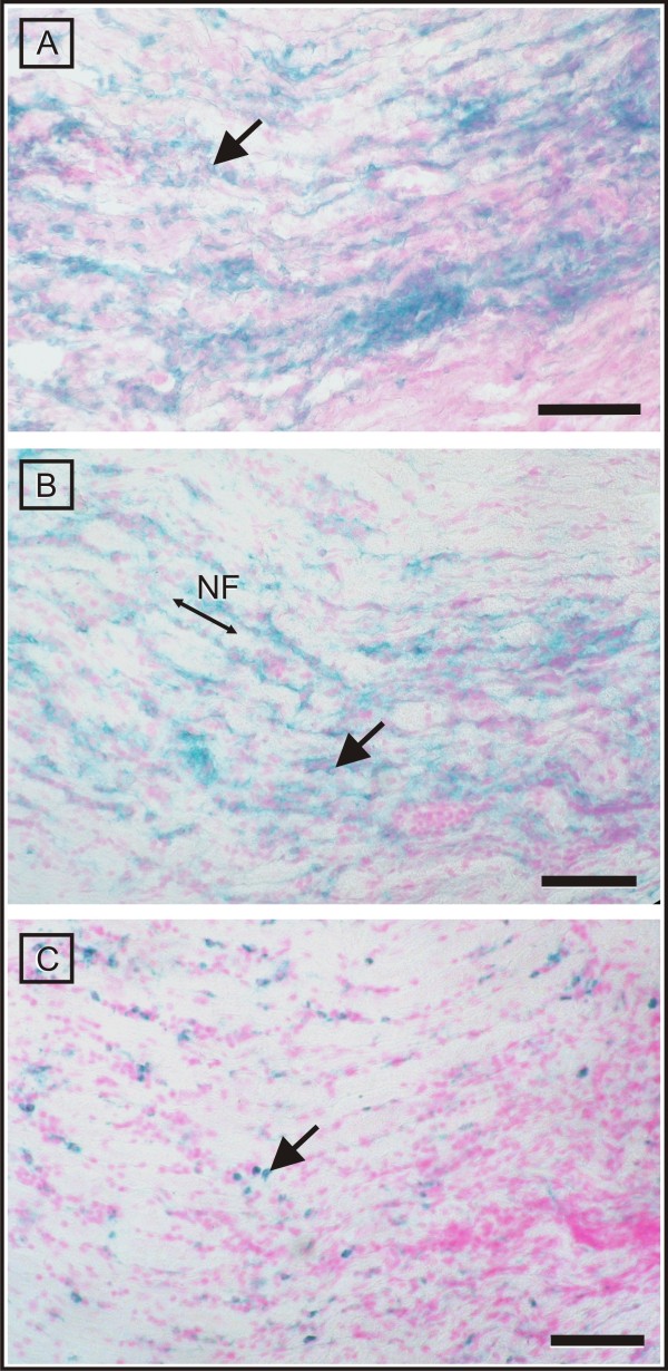
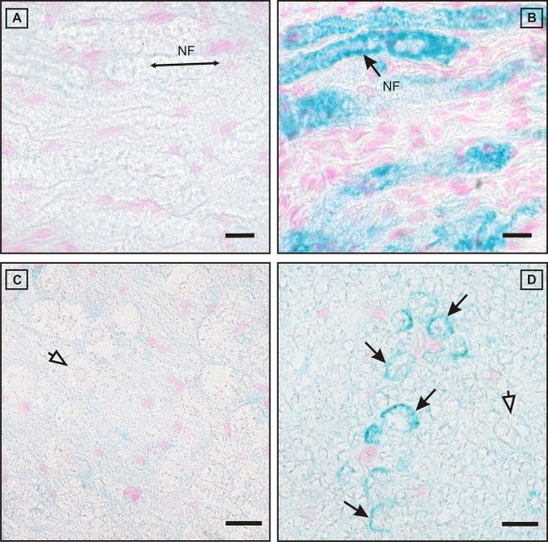
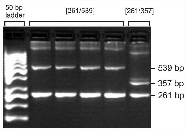
Results: Spontaneous paresis of White Leghorns is caused by cell-mediated, inflammatory demyelination affecting multiple cranial and spinal nerves and nerve roots with a proximodistal tapering. Clinical manifestation coincides with the employment of humoral immune mechanisms, enrolling plasma cell recruitment, deposition of myelin-bound IgG and antibody-dependent macrophageal myelin-stripping. Disease development was significantly linked to a 539 bp microsatellite in MHC locus LEI0258. An aetiological role for MDV was excluded.

Conclusions: The paretic phase of avian inflammatory demyelinating polyradiculoneuritis immunobiologically resembles the late-acute disease stages of human acute inflammatory demyelinating polyneuropathy, and is characterised by a Th1-to-Th2 shift.


Figures

Figure 1
Figure 1
Gross findings in AvIDP-affected chickens. Compared to controls (A, C) a marked thickening of craniospinal nerve roots was recognized in all affected birds (B, D). A, B: Inspection of the ventral brain surface reveals severely enlarged maxillary (CN Vmax) and ophthalmic (CN Vopht) nerve branches. The oculomotor (CN III) and abducens (CN VI) nerve are also thickened to a lesser extend, and show a mild greyish discoloration. C, D: The dorsal view at the cervical intumescence between C13 and Th1 indicates severe enlargement of the dorsal rootlets (arrows) and the associated dorsal root ganglia (DRG). Affected ganglia (b) measure up to fourfold the diameters of unaffected control nerves (a). A, B: EB = eye ball; NO = optic nerve; P = Pons; scale bar = 1.0 cm. C, D: SC: spinal cord; scale bar 0.5 cm.
Figure 2
Figure 2
CNS-PNS transition as the border of inflammation. The arrows point to the CNS - PNS boundary of the spinal roots. Despite the severe involvement of both dorsal (DR) and ventral (VR) nerve roots, the inflammatory infiltration spares the adjacent central white matter and does not pass the PNS-CNS transition. Scale bar = 0.5 mm.
Figure 3
Figure 3
CNS-PNS transition at the dorsal nerve root as the border of inflammation. On closer inspection, the dorsal root (DR) of an affected animal (B) shows a mononuclear infiltration in comparison to a control chicken (A). The arrows point to the CNS - PNS boundary. Note that the adjacent white (WM) and grey matters (GM) are not affected. Scale bar = 1 mm.
Figure 4
Figure 4
CNS-PNS transition at the ventral nerve root as the border of inflammation. The ventral nerve root (VR) of an affected animal (B) presents with a severe mononuclear infiltration and thickening in comparison to a control chicken (A). The arrows point to the CNS - PNS boundary. Again, the inflammatory infiltration spares the adjacent central white (WM) and grey matters (GM) and does not pass the PNS-CNS transition. Scale bar = 1 mm.
Figure 5
Figure 5
Immunohistochemical illustration of Ricinus communis agglutinin-1 (RCA-1) -positive microglial cells in the white matter of spinal cord. The RCA-1-positive microglial cells (arrowheads) in both healthy (A) and AvIDP-affected (B) chicken show incospicuous ramified microglial cells. Activated microglial cells or an increased number were not observed. A, B: asterisk = endothelium (also stains RCA-1 positive); scale bar = 250 μm.
Figure 6
Figure 6
AvIDP: typical histological PNS findings. The histological appearance of a healthy mixed fascicular nerve is displayed in figure A. Its endoneurium (E) contains large (LMF) and small (SMF) myelinated fibres that are confined by a peripheral rim of blue (azurophilic) compacted myelin. B: AvIDP leads to a breakdown of the myelin sheath while leaving the axons mainly untouched. Concomitantly with the myelin loss (arrow), an expansion of the eosinophilic extracellular matrix of the endoneurial sheath develops which results in a decrease of nerve fibre density. A, B: P = perineurium; scale bar = 100 μm.
Figure 7
Figure 7
Macrophage-mediated myelin stripping. A: Upon invasion of the Schwann cell tubes, macrophage processes (arrows) split the outer mesaxon in order to gain entry to the myelin spiral. B: They invade the myelin following the intraperiod line (arrow). Widening propagates centripetally towards the inner mesaxon. B: MS = myelin sheath; Scale bar = 2 μm.
Figure 8
Figure 8
Ongoing demyelination/remyelination. A: Demyelination is accompanied by proliferation of Schwann cells (SC), formation of supernumerary processes, and a significant expansion of the collagenous inner endoneurial sheath. Schwann cells show no sign of degeneration. B: Inappropriately thin myelin sheaths and redundant Schwann cells are consistent with ongoing remyelination. Apart from macrophages, the endoneurium of affected nerves shows significant infiltration by plasma cells and lymphocytes (LC). A: FB = fibroblast; Scale bars = 1 μm (A), 250 nm (B).
Figure 9
Figure 9
Immunohistochemical evidence of infiltration by T-cells (CD3+), B-cells (chB6+) and macrophages (Kul01+). Immunophenotyping identified the majority of infiltrating lymphocytes as being CD3-positive T-cells (A). They show a characteristic multifocal distribution pattern. With similar spatial characteristics, chB6-positive B-cells comprise the second largest fraction of infiltrating cells (B). Compared to the lymphocytes, the density of Kul01-positive macrophages appears much lower (C). The arrows point to immunopositive cells. All sections were prepared from the same region of the sciatic nerve. The orientation of the nerve fibres (NF) is indicated. Scale bar = 35 μm.
Figure 10
Figure 10
Deposition of IgG within the myelin sheath of affected fibres. Longitudinal (A) and cross (C) sections of unaffected samples show immunonegative nerve fibres (NF) throughout (empty arrow), whereas inflamed specimens reveal a significant intramyelinic IgG-deposition (B, D; black arrows) in many fibres within (B) or even outside (D) of significantly infiltrated foci. The longitudinal orientation of the nerve fibers is indicated by the double-headed arrow (A). Scale bars = 10 μm (A, B), 15 μm (C, D).
Figure 11
Figure 11
Leukocytes in spinal ganglia from diseased birds consist of different subpopulations. For flow cytometric analysis, single cell suspensions from spinal ganglia of diseased birds were stained with a Pan-Leukocyte marker (CD45) and antibodies for T cells (CD3), cytotoxic T cells (CD8), T helper cells (CD4), T cell receptor types TCR1, TCR2 and TCR3 (TCRγδ, TCRα/vβ1, TCRα/vβ2), B cells (AV20), macrophages (Kul01) and MHC class II (2G11). (A) Gating was first performed on cells with leukocyte scatter characteristics, secondly on CD45+ cells before the proportion of different lymphocyte subpopulations of all CD45+ leukocytes was determined. (B) Data expressed as mean ± SD for different subpopulations for five birds analysed.
Figure 12
Figure 12
Relative Gene expression in spinal ganglia of chicken. The mRNA abundance of candidate genes using quantitative RT-PCR was analysed in ganglia of affected and non-affected chickens of the same age, originated from the same flock. Gene expression, measured using SYBR Green primer assays and Ct values, were normalised against 18S rRNA (= dCT). Data shown as mean ± SD for 40-dCT values for eight animals per group (** p ≤ 0.01; * p ≤ 0.05).
Figure 13
Figure 13
Fragment length polymorphism of LEI0258. Blood group genotyping revealed an association between AvIDP and MHC haplotype with a fragment size of 539 bp. The individual with genotype [261/357] was healthy.

Similar articles

Cited by

References

    1. Yu RK, Usuki S, Ariga T. Ganglioside molecular mimicry and its pathological roles in Guillain-Barre syndrome and related diseases. Infect Immun. 2006;74:6517–6527. doi: 10.1128/IAI.00967-06. - DOI - PMC - PubMed
    1. Hughes RA, Hadden RD, Gregson NA, Smith KJ. Pathogenesis of Guillain-Barre syndrome. J Neuroimmunol. 1999;100:74–97. doi: 10.1016/S0165-5728(99)00195-2. - DOI - PubMed
    1. Latov N. Diagnosis of CIDP. Neurology. 2002;59:S2–6. - PubMed
    1. McCombe PA, Pollard JD, McLeod JG. Chronic inflammatory demyelinating polyradiculoneuropathy. A clinical and electrophysiological study of 92 cases. Brain. 1987;110(Pt 6):1617–1630. doi: 10.1093/brain/110.6.1617. - DOI - PubMed
    1. Hadden RD, Hughes RA. Management of inflammatory neuropathies. J Neurol Neurosurg Psychiatry. 2003;74(Suppl 2):ii9–ii14. - PMC - PubMed

Publication types

MeSH terms