

Prediction of clinical toxicity in locally advanced head and neck cancer patients by radio-induced apoptosis in peripheral blood lymphocytes (PBLs)
- PMID: 20109191
- PMCID: PMC2827476
- DOI: 10.1186/1748-717X-5-4
Prediction of clinical toxicity in locally advanced head and neck cancer patients by radio-induced apoptosis in peripheral blood lymphocytes (PBLs)
Abstract
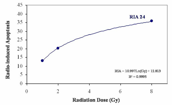
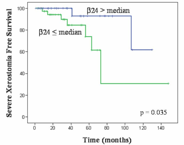
Head and neck cancer is treated mainly by surgery and radiotherapy. Normal tissue toxicity due to x-ray exposure is a limiting factor for treatment success. Many efforts have been employed to develop predictive tests applied to clinical practice. Determination of lymphocyte radio-sensitivity by radio-induced apoptosis arises as a possible method to predict tissue toxicity due to radiotherapy. The aim of the present study was to analyze radio-induced apoptosis of peripheral blood lymphocytes in head and neck cancer patients and to explore their role in predicting radiation induced toxicity. Seventy nine consecutive patients suffering from head and neck cancer, diagnosed and treated in our institution, were included in the study. Toxicity was evaluated using the Radiation Therapy Oncology Group scale. Peripheral blood lymphocytes were isolated and irradiated at 0, 1, 2 and 8 Gy during 24 hours. Apoptosis was measured by flow cytometry using annexin V/propidium iodide. Lymphocytes were marked with CD45 APC-conjugated monoclonal antibody. Radiation-induced apoptosis increased in order to radiation dose and fitted to a semi logarithmic model defined by two constants: alpha and beta. Alpha, as the origin of the curve in the Y axis determining the percentage of spontaneous cell death, and beta, as the slope of the curve determining the percentage of cell death induced at a determined radiation dose, were obtained. beta value was statistically associated to normal tissue toxicity in terms of severe xerostomia, as higher levels of apoptosis were observed in patients with low toxicity (p = 0.035; Exp(B) 0.224, I.C.95% (0.060-0.904)). These data agree with our previous results and suggest that it is possible to estimate the radiosensitivity of peripheral blood lymphocytes from patients determining the radiation induced apoptosis with annexin V/propidium iodide staining. beta values observed define an individual radiosensitivity profile that could predict late toxicity due to radiotherapy in locally advanced head and neck cancer patients. Anyhow, prospective studies with different cancer types and higher number of patients are needed to validate these results.
Figures
References
-
- Johansson S, Svensson H, Denekamp J. Timescale of evolution of late radiation injury after postoperative radiotherapy of breast cancer patients. Int J Radiat Oncol Biol Phys. 2000;48:745–750. - PubMed
-
- Fernet M, Hall J. Predictive markers for normal tissue reactions: fantasy or reality? Cancer Radiother. 2008;12:614–618. - PubMed
-
- Buchholz TA. Finding our sensitive patients. Int J Radiat Oncol Biol Phys. 1999;45:547–548. - PubMed
-
- Hennequin C, Quero L, Favaudon V. [Determinants and predictive factors of tumour radiosensitivity] Cancer Radiother. 2008;12:3–13. - PubMed
Publication types
MeSH terms
LinkOut - more resources
Full Text Sources
Medical
Research Materials
Miscellaneous
