. 2010 Jan 28:7:8.
doi: 10.1186/1742-2094-7-8.

Progressive changes in microglia and macrophages in spinal cord and peripheral nerve in the transgenic rat model of amyotrophic lateral sclerosis

Affiliations

Progressive changes in microglia and macrophages in spinal cord and peripheral nerve in the transgenic rat model of amyotrophic lateral sclerosis

David J Graber et al. J Neuroinflammation. .

Abstract

Background: The role of neuroinflammation in motor neuron death of amyotrophic lateral sclerosis (ALS) is unclear. The human mutant superoxide dismutase-1 (hmSOD1)-expressing murine transgenic model of ALS has provided some insight into changes in microglia activity during disease progression. The purpose of this study was to gain further knowledge by characterizing the immunological changes during disease progression in the spinal cord and peripheral nerve using the more recently developed hmSOD1 rat transgenic model of ALS.

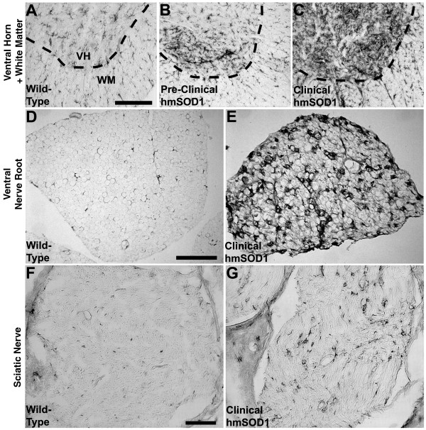
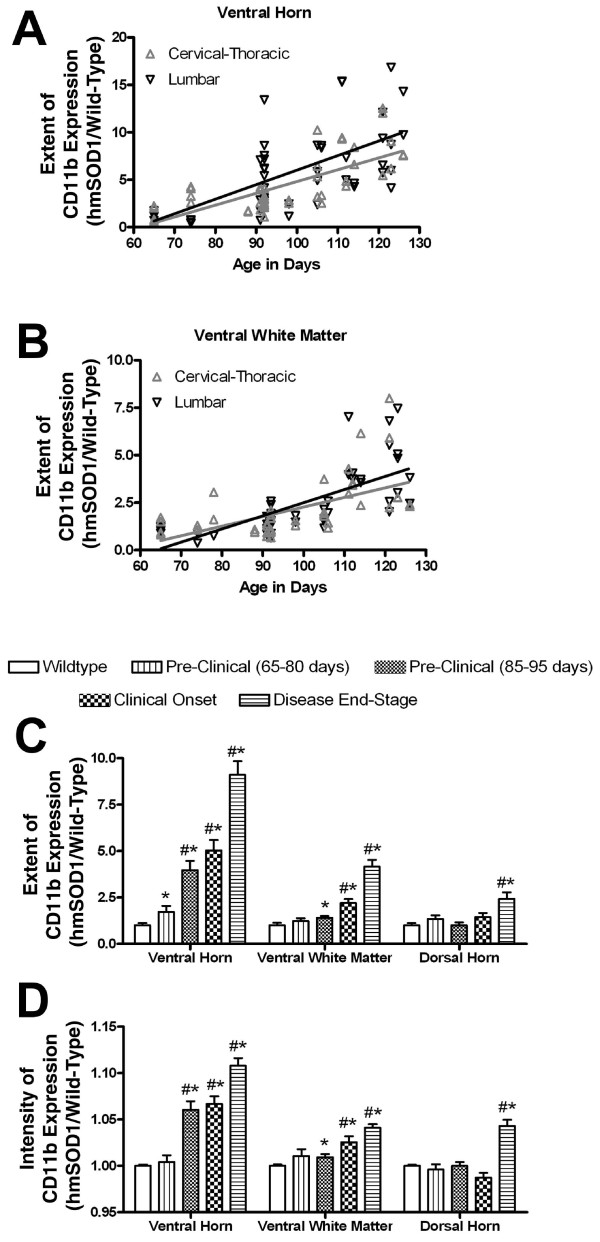
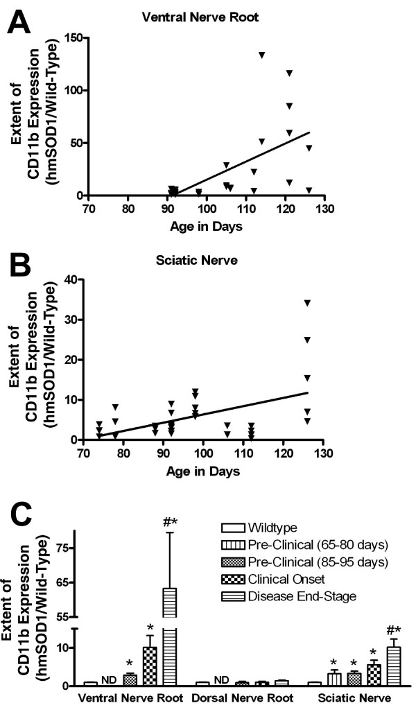
Methods: Using immunohistochemistry, the extent and intensity of tissue CD11b expression in spinal cord, lumbar nerve roots, and sciatic nerve were evaluated in hmSOD1 rats that were pre-clinical, at clinical onset, and near disease end-stage. Changes in CD11b expression were compared to the detection of MHC class II and CD68 microglial activation markers in the ventral horn of the spinal cord, as well as to the changes in astrocytic GFAP expression.

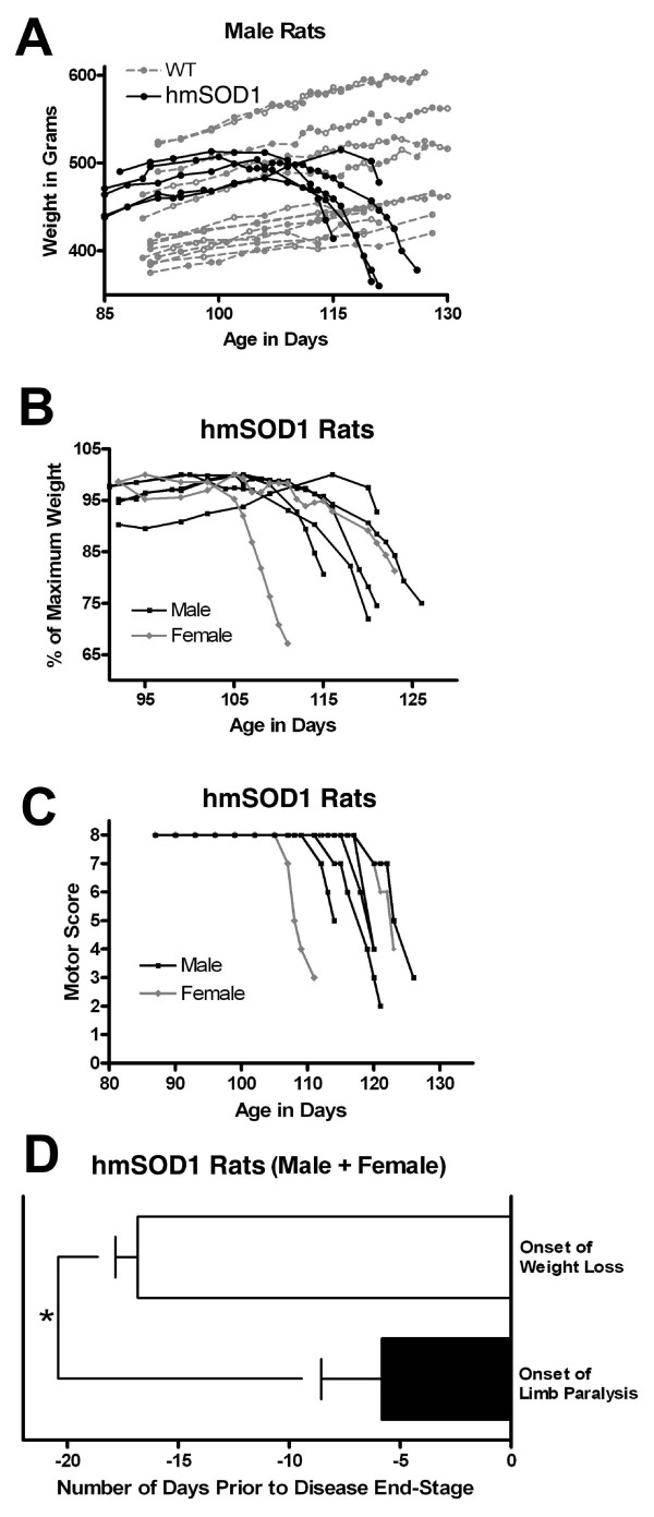
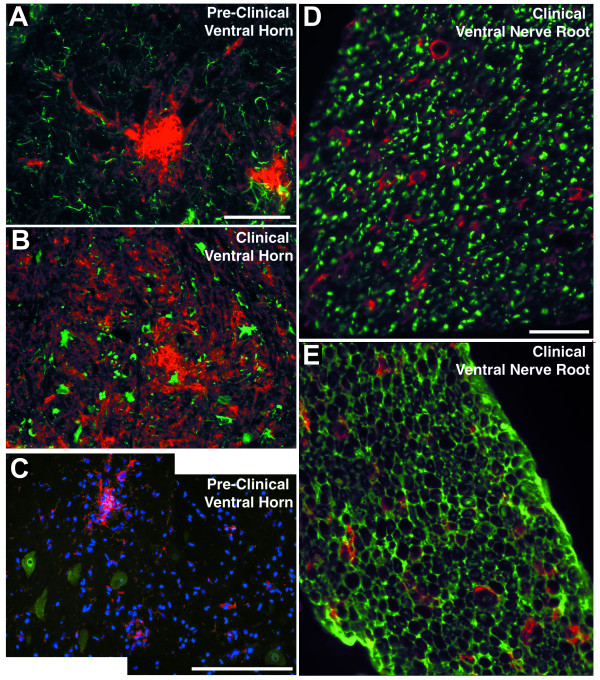
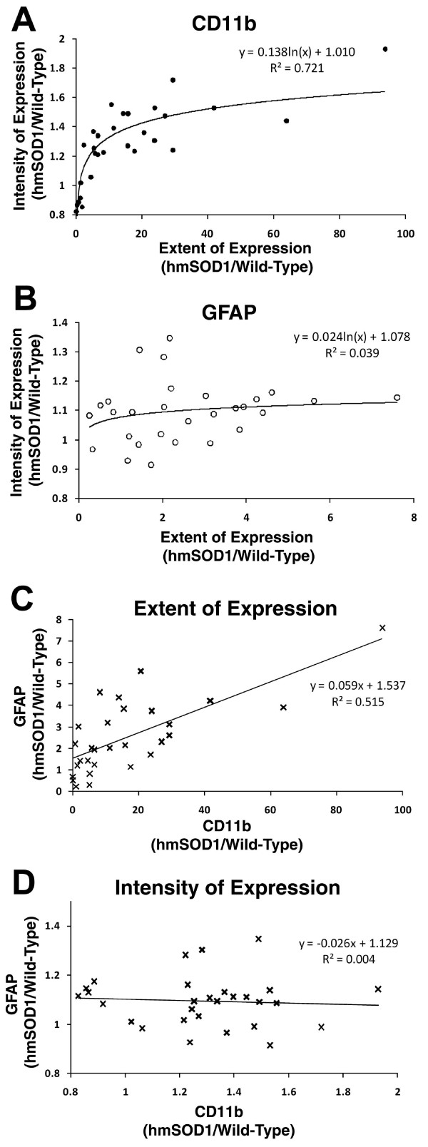
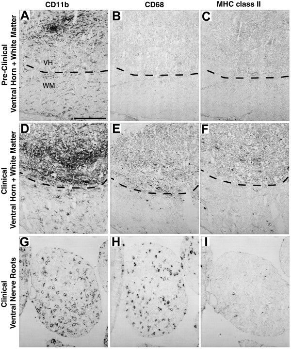
Results: Our study reveals an accumulation of microglia/macrophages both in the spinal cord and peripheral nerve prior to clinical onset based on CD11b tissue expression. The microglia formed focal aggregates in the ventral horn and became more widespread as the disease progressed. Hypertrophic astrocytes were not prominent in the ventral horn until after clinical onset, and the enhancement of GFAP did not have a strong correlation to increased CD11b expression. Detection of MHC class II and CD68 expression was found in the ventral horn only after clinical onset. The macrophages in the ventral nerve root and sciatic nerve of hmSOD1 rats were observed encircling axons.

Conclusions: These findings describe for the first time in the hmSOD1 rat transgenic model of ALS that enhancement of microglia/macrophage activity occurs pre-clinically both in the peripheral nerve and in the spinal cord. CD11b expression is shown to be a superior indicator for early immunological changes compared to other microglia activation markers and astrogliosis. Furthermore, we suggest that the early activity of microglia/macrophages is involved in the early phase of motor neuron degeneration and propose that studies involving immunomodulation in hmSOD1transgenic models need to consider effects on macrophages in peripheral nerves as well as to microglia in the spinal cord.


Figures

Figure 1
Figure 1
Clinical onset and progression of disease. Weight loss and limb motor abnormalities in hmSOD1 transgenic rats as a function of age. (A) Weight in grams of male hmSOD1 (black) and wild-type littermates (gray). (B) Weight expressed as percent of maximum recorded weight in male (black) and female (gray) hmSOD1 rats. (C) Limb paralysis motor score of male (black) and female (gray) hmSOD1 rats. See Table 1 for scoring criteria. (D) Average time between age of onset for weight loss and limb paralysis relative to disease end-stage in hmSOD1 rats. *, significantly different (P < 0.01, paired T test).
Figure 2
Figure 2
CD11b-labeld microglia/macrophages in spinal cord and nerve. Microglia or macrophages in the ventral spinal cord, ventral nerve roots, and sciatic nerve as assessed by CD11b histological expression. (A) CD11b-labeled microglia are evenly distributed in the ventral horn (VH, above dashed line) and in the ventral white matter (WM, below dashed line) of wild-type rats. (B) In pre-clinical hmSOD1 rats, focal increases in CD11b expression are often observed in the ventral horn. (C) After clinical onset, the extent of CD11b expression increases in ventral horn and to a lesser degree in the adjacent ventral white matter. Scale bar in panel A represents 200 μm for A-C. (D) There are very few CD11b-labeled resident macrophages in ventral nerve roots of wild-type rats. (E) After clinical onset in hmSOD1 rats, there is an increase in macrophages in the ventral nerve root. Scale bar in panel D represents 100 μm for D and E. (F) Very few CD11b-labeled resident macrophages are found in sciatic nerve of wild-type rats. (G) After clinical onset in hmSOD1 rats, there is an increase in macrophages in the sciatic nerve. Scale bar in panel F represents 100 μm for F and G.
Figure 3
Figure 3
Regional changes in CD11b expression in spinal cord. Changes in the extent and intensity of DAB-stained CD11b expression in spinal cord of hmSOD1 rats as a function of age or disease stage. (A, B) The extent of CD11b expression relative to age-matched wild-type levels is expressed as a function of age. Linear regression slopes in the ventral horns of 0.12 ± 0.018 at the cervical-thoracic level and 0.15 ± 0.028 at the lumbar level (A). Linear regression slopes in the ventrolateral white matter are 0.05 ± 0.011 at the cervical-thoracic level and 0.069 ± 0.0107 at the lumbar level (B). (C, D) The average extent (C) and intensity (D) of CD11b expression relative to wild-type levels in spinal cord regions expressed as a function of disease stage. Bars represent average + SEM. #, significantly different compared to wild-type levels (P < 5 × 10⁻⁵, Neuman-Keuls test). *, significantly different compared to levels from only age-matched wild-type rats (P < 0.05, unpaired T test).
Figure 4
Figure 4
Changes in CD11b expression in nerve roots and sciatic nerve. Changes in the extent of DAB-stained CD11b expression in lumbar nerve roots and sciatic nerves of hmSOD1 rats. (A,B) The extent of CD11b expression relative to age-matched wild-type levels is expressed as a function of age. Linear regression slopes are 1.7 ± 0.45 in the ventral nerve roots (A) and 0.21 ± 0.063 in the sciatic nerve (B). (C) The average extent of CD11b expression relative to wild-type levels is expressed as a function of disease stage. Bars represent average + SEM. ND, not determined. #, significantly different compared to wild-type levels (P < 0.0005, Neuman-Keuls test). *, significantly different compared to levels from only age-matched wild-type rats (P < 0.05, unpaired T test).
Figure 5
Figure 5
Microglia/macrophages with neurons, astrocytes, and Schwann cells. Dual immunofluorescence of microglia in the ventral horn and macrophages in the ventral nerve root of hmSOD1 rat. (A,B) GFAP-labeled astrocytes (green) and CD11b-labeled microglia (red) in a ventral horn at pre-clinical stage (A) and after clinical onset (B). Scale bar in panel A represents 100 μm for A and B. (C) Composite image showing Iba1-labeled microglia (red) near NeuN-labeled motor neurons (green) in a pre-clinical rat. Scale bar in panel C represents 200 μm. (D,E) CD11b-labeled macrophages (red) relative to βIII-tubulin-labeled axons (D, green) or S100-labeled Schwann cells in ventral nerve root after clinical onset (E, green). Scale bar in D represents 100 μm for D and E.
Figure 6
Figure 6
Correlation between CD11b and GFAP expression in ventral horn. Changes in GFAP and CD11b in lumbar ventral horn detected with dual immunofluorescence throughout disease progression. (A,B) The extent relative to intensity of CD11b (A) and GFAP (B) expression. (C) The extent of expression of CD11b relative to GFAP expression. (D) The tissue intensity of CD11b relative to GFAP expression.
Figure 7
Figure 7
Expression of CD11b, CD68, and MHC class II in spinal cord and ventral nerve root. Expression of CD11b, CD68, and MHC II in adjacent sections of ventral spinal cord and ventral nerve root of hmSOD1 rats. (A-C) In pre-clinical stage ventral horn, a cluster of CD11b-labeled microglia (A) in the ventral horn (VH, above dashed line) does not co-localize with CD68 (B) or MHC II (C). (D-F) In the ventral horn after clinical onset, CD11b expression (D) is more widespread and both CD68 (E) and MHC II (F) expression are detected. (G-I) In ventral nerve root after clinical onset, both CD11b- (G) and CD68-labeled (H) cells are increased, but MHC II-labeled cells (I) remain sparse. Scale bar in panel A represents 200 μm for all panels.

Similar articles

Cited by

References

    1. Kawamata T, Akiyama H, Yamada T, McGeer PL. Immunologic reactions in amyotrophic lateral sclerosis brain and spinal cord tissue. Am J Pathol. 1992;140:691–707. - PMC - PubMed
    1. Ince PG, Shaw PJ, Slade JY, Jones C, Hudgson P. Familial amyotrophic lateral sclerosis with a mutation in exon 4 of the Cu/Zn superoxide dismutase gene: pathological and immunocytochemical changes. Acta Neuropathol. 1996;92:395–403. doi: 10.1007/s004010050535. - DOI - PubMed
    1. Hall ED, Oostveen JA, Gurney ME. Relationship of microglial and astrocytic activation to disease onset and progression in a transgenic model of familial ALS. Glia. 1998;23:249–56. doi: 10.1002/(SICI)1098-1136(199807)23:3<249::AID-GLIA7>3.0.CO;2-#. - DOI - PubMed
    1. Magnus T, Carmen J, Deleon J, Xue H, Pardo AC, Lepore AC, Mattson MP, Rao MS, Maragakis NJ. Adult glial precursor proliferation in mutant SOD1(G93A) mice. Glia. 2008;56:200–8. doi: 10.1002/glia.20604. - DOI - PubMed
    1. Fendrick SE, Xue QS, Streit WJ. Formation of multinucleated giant cells and microglial degeneration in rats expressing a mutant Cu/Zn superoxide dismutase gene. J Neuroinflammation. 2007;4:9. doi: 10.1186/1742-2094-4-9. - DOI - PMC - PubMed

Publication types

MeSH terms