. 2010 Jan 29:6:8.
doi: 10.1186/1744-8069-6-8.

Trigeminal injury causes kappa opioid-dependent allodynic, glial and immune cell responses in mice

Affiliations

Trigeminal injury causes kappa opioid-dependent allodynic, glial and immune cell responses in mice

Megumi Aita et al. Mol Pain. .

Abstract

Background: The dynorphin-kappa opioid receptor (KOR) system regulates glial proliferation after sciatic nerve injury. Here, we investigated its role in cell proliferation following partial ligation of infraorbital nerve (pIONL), a model for trigeminal neuropathic pain. Mechanical allodynia was enhanced in KOR gene deleted mice (KOR-/-) compared to wild type mice. Using bromodeoxyuridine (BrdU) as a mitotic marker, we assessed cell proliferation in three different areas of the trigeminal afferent pathway: trigeminal nucleus principalis (Vp), trigeminal root entry zone (TREZ), and trigeminal ganglion (TG).

Results: In KOR-/- mice or norBNI-treated mice, the number of proliferating cells in the Vp was significantly less than in WT mice, whereas cell proliferation was enhanced in TREZ and TG. The majority of the proliferating cells were nestin positive stem cells or CD11b positive microglia in the Vp and macrophages in the TG. GFAP-positive astrocytes made a clear borderline between the CNS and the PNS in TREZ, and phosphorylated KOR staining (KOR-p) was detectable only in the astrocytes in CNS in WT mice but not in KOR-/- or norBNI-treated mice.

Conclusions: These results show that kappa opioid receptor system has different effects after pIONL in CNS and PNS: KOR activation promotes CNS astrocytosis and microglial or stem cell proliferation but inhibits macrophage proliferation in PNS. The trigeminal central root has a key role in the etiology and treatment of trigeminal neuralgia, and these newly identified responses may provide new targets for developing pain therapies.


Figures

Figure 1
Figure 1
(A) Response thresholds of ipsilateral whisker pad to usually innocuous tactile (von Frey Hair) stimuli in WT sham, WT pIONL, KOR-/- sham, and KOR-/- pIONL mice during 25 days after partial infraorbital nerve ligation. After pIONL, both WT and KOR-/- mice developed allodynic response as evident from the decreased thresholds to tactile stimulation compared with WT and KOR-/- mice that received sham-ligation surgeries. One week after pIONL, WT mice with pIONL began to show the increase of the mechanical threshold, whereas KOR-/- pIONL group showed much slower recovery and significantly greater allodynic response than WT pIONL group. (B)On the 25th day after surgery, we injected the kappa opioid receptor agonist U50,488 intraperitoneally at 10 mg/kg in each group of mice. The von Frey hair force returned to baseline in WT pIONL group, but not in KOR-/- pIONL group. U50,488 injection did not affect the mechanical threshold in sham operated groups. Asterisks indicate significant decreases in the threshold to stimuli by the von Frey hair applied to ipsilateral side of whisker pad in KOR-/- pIONL group compared to WT pIONL group (p < 0.05, ANOVA followed by Student-Newman test). Data are presented as the mean ± SEM von Frey hair threshold in grams; n = 8 per time point).
Figure 2
Figure 2
Face rubbing after pIONL. The total time for isolated face-rubbing episode during a 15 min observation period. The spontaneous face-rubbing behavior was recorded for 15 minutes on preoperative day 1 (baseline), and from postoperative day1 to day 6. The duration of face-rubbing behavior was elevated for both WT and KOR-/- pIONL groups on the first day after surgery compared to the baseline and sham operated groups. KOR-/- pIONL group showed longer duration of face-rubbing behavior than that of WT pIONL group on the postoperative day 1 (p < 0.05, ANOVA followed by Student-Newman test, n = 8 per group). The duration of face-rubbing behavior peaked at postoperative day 1 and this increased facial rubbing gradually recovered to the basal level.
Figure 3
Figure 3
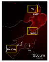
Orientation of VP, TREZ, TG. Key areas for our analyses are shown here: (1) the trigeminal principle nucleus (Vp), (2) The root entry zone (TREZ) where central glia are in the CNS side (GFAP, red), and Schwann cells on the ganglion side, and (3) trigeminal ganglion (TG). The cell bodies for infraorbital nerve (ION) occur in the distal parts of the combined V1/V2 but not in V3. Yellow boxes show sites for quantification.
Figure 4
Figure 4
GFAP immunoreactivity in the TREZ at day 8 after pIONL. (A), GFAP-IR in sham operated WT mice. (B, C), pIONL induced slight increase in the intensity of GFAP staining in the ipsilateral TREZ of WT mice but not the contralateral side. (D, E), KOR-/- mice or mice pre-treated with norBNI did not show significant upregulation of GFAP immunoreactivity in the ipsilateral TREZ, and this was the same intensity as in the contralateral TREZ of WT pIONL mice (B) or WT sham operated mice(A). (F), Mean ± SEM pixel intensity of GFAP immunoreactivity in TREZ at day 8 after pIONL. (*p < 0.001) Scale bars: A-E, 50 μm
Figure 5
Figure 5
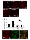
KOR activation in TREZ at day 8 after pIONL. (A), KOR-p-IR in sham operated WT mice. (B, C), increased KOR-p staining was seen within the root entry zone on the ipsilateral side compared to contralateral side following pIONL in WT mice. KOR activation was blocked by the deletion of kappa opioid receptor or norBNI treatment (D, E). (F), Mean ± SEM pixel intensity of KOR-p intensity in TREZ at day 8 after pIONL. (G-I), Double immunohistochemistry for KOR-p and GFAP in the TREZ at day 8 after pIONL. KOR-p staining is completely overlapped with GFAP staining. (*p < 0.001) Scale bars: A-E, 30 μm; G-I, 50 μm.
Figure 6
Figure 6
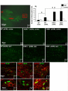
Cell proliferation central trigeminal root at day 8 after pIONL. (A) Data came from Vp (the area shown in red box). (B, C, F), BrdU positive cells are markedly increased in the ipsilateral Vp inWT pIONL compare to contralateral side. (B-H), in KOR-/- and NBNI pre-treated pIONL mice, the number of BrdU positive cells significantly decreased compared to WT pIONL mice. (I-N), Different cellular markers (GFAP for satellite cells, CD11b for microglia, nestin for neuronal stem cell) were used to identify the phenotypes of dividing cells. The majority of BrdU positive cells were double labeled with nestin and CD11b. Only few BrdU positive nuclei were double labeled with GFAP. (*p < 0.001) Scale bars: A, 250 μm, C-H, 50 μm, I-N, 30 μm.
Figure 7
Figure 7
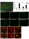
Cell proliferation in TREZ 8 d after pIONL. (A) Data came from TREZ (the area shown in red box). (B, C, F), the number of BrdU positive nuclei increased in both ipsilateral and contralateral TREZ following pIONL in WT compared to sham operated mice. (B-H), in the ipsilateral TREZ, KOR-/- and norBNI treated mice showed BrdU positive nuclei more than twice as much as that in WT mice. Similar to the result from Vp, the majority of BrdU positive nuclei was double-labeled with CD11b or Nestin in the CNS side of TREZ. Neither GFAP or S100beta-IR was double labeled with BrdU-IR (I-P). (*p < 0.001) Scale bars: A, 250 μm, C-H, 50 μm, I-P, 30 μm.
Figure 8
Figure 8
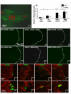
Cell proliferation in ION region in the trigeminal ganglion at day 8 after pIONL. (A) Data came from ION region in TG (the area shown in red box). (B, C, F), pIONL induced marked upregulation of BrdU positive cells in the ipsilateral side of ION region in WT mice. (B-H), in KOR-/- and norBNI pre-treated mice, the number of BrdU positive cells significantly increased compared to WT pIONL mice. (I-N) Different cellular markers (GFAP for satellite cells, S100beta for Schwann cells, CD11b for macrophage) were used to identify the phenotypes of dividing cells. Only rare cells were double labeled for BrdU and GFAP or S100beta. The majority of BrdU positive cells were CD11b positive macrophage. (*p < 0.001). Scale bars: A, 250 μm, C-H, 50 μm, I-N, 30 μm.

Similar articles

Cited by

References

    1. Cheshire WP. Trigeminal neuralgia: for one nerve a multitude of treatments. Expert Rev Neurother. 2007;7:1565–1579. doi: 10.1586/14737175.7.11.1565. - DOI - PubMed
    1. Xu M, Aita M, Chavkin C. Partial infraorbital nerve ligation as a model of trigeminal nerve injury in the mouse: behavioral, neural, and glial reactions. J Pain. 2008;9:1036–1048. doi: 10.1016/j.jpain.2008.06.006. - DOI - PMC - PubMed
    1. Xu M, Petraschka M, McLaughlin JP, Westenbroek RE, Caron MG, Lefkowitz RJ, Czyzyk TA, Pintar JE, Terman GW, Chavkin C. Neuropathic pain activates the endogenous kappa opioid system in mouse spinal cord and induces opioid receptor tolerance. J Neurosci. 2004;24:4576–4584. doi: 10.1523/JNEUROSCI.5552-03.2004. - DOI - PMC - PubMed
    1. Toma J, McPhail LT, Ramer MS. Comparative postnatal development of spinal, trigeminal and vagal sensory root entry zones. Int J Devel Neurosci. 2006;24:373–388. doi: 10.1016/j.ijdevneu.2006.06.001. - DOI - PubMed
    1. Jannetta PJ, Abbasy M, Maroon JC, Ramos FM, Albin MS. Etiology and definitive microsurgical treatment of hemifacial spasm. Operative techniques and results in 47 patients. J Neurosurg. 1977;47:321–8. doi: 10.3171/jns.1977.47.3.0321. - DOI - PubMed

Publication types

MeSH terms