. 2010 May 15;316(9):1600-9.
doi: 10.1016/j.yexcr.2010.01.021. Epub 2010 Jan 25.

Downregulation of FAK-related non-kinase mediates the migratory phenotype of human fibrotic lung fibroblasts

Affiliations

Downregulation of FAK-related non-kinase mediates the migratory phenotype of human fibrotic lung fibroblasts

Guo-qiang Cai et al. Exp Cell Res. .

Abstract

Fibroblast migration plays an important role in the normal wound healing process; however, dysregulated cell migration may contribute to the progressive formation of fibrotic lesions in the diseased condition. To examine the role of focal-adhesion-kinase (FAK)-related non-kinase (FRNK) in regulation of fibrotic lung fibroblast migration, we examined cell migration, FRNK expression, and activation of focal adhesion kinase (FAK) and Rho GTPase (Rho and Rac) in primary lung fibroblasts derived from both idiopathic pulmonary fibrosis (IPF) patients and normal human controls. Fibrotic (IPF) lung fibroblasts have increased cell migration when compared to control human lung fibroblasts. FRNK expression is significantly reduced in IPF lung fibroblasts, while activation of FAK, Rho and Rac is increased in IPF lung fibroblasts. Endogenous FRNK expression is inversely correlated with FAK activation and cell migration rate in IPF lung fibroblasts. Forced exogenous FRNK expression abrogates the increased cell migration, and blocked the activation of FAK and Rho GTPase (Rho and Rac), in IPF lung fibroblasts. These data for the first time provide evidence that downregulation of endogenous FRNK plays a role in promoting cell migration through FAK and Rho GTPase in fibrotic IPF lung fibroblasts.


Figures

Fig. 1
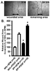
Fig. 1. IPF lung fibroblasts demonstrate enhanced cell migration under basal (serum-free) and PDGF-BB-stimulated conditions
(A): Serum-starved normal human lung fibroblasts were wounded, and the monolayer wound area was monitored for 24 hours at 37°C. Representative digital images at 0-hour and 24-hour are shown. (B): Migration of normal human lung fibroblasts (NHL) and IPF lung fibroblasts (IPF) were examined as in Panel A. NHL and IPF lung fibroblasts were wounded and treated without or with PDGF-BB (4 ng/ml) in serum-free media (SFM) with 1% BSA as in Panel A. Data were pooled from six NHL and ten IPF human lung fibroblast lines, and plotted (mean + SE) as % of wound area covered over 24 hours relative to normal human lung fibroblasts (NHL). The wound area covered over 24 hours in 19LU normal human lung fibroblasts was defined as 100%. The black bars represent the IPF lung fibroblasts. * represents p < 0.01 relative to NHL group in SFM. # represents p < 0.01 relative to NHL group treated with PDGF-BB.
Fig. 2
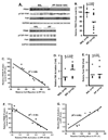
Fig. 2. FRNK protein level is decreased and FAK activation is increased in IPF lung fibroblasts
(A): Normal human lung fibroblasts (NHL, n = 6), and IPF lung fibroblasts (IPF, n = 10) were examined. IPF lung fibroblasts with an increased cell migration rate were presented at the Top-Panel (IPF, faster rate, n = 6). IPF lung fibroblasts (n = 4) having a similar cell migration rate as NHL were presented in the Bottom-Panel. Endogenous FRNK expression was examined by immunoprecipitation (IP) of equivalent amount of whole cell detergent lysates, and Western blotted with antibody directed toward FRNK. Equivalent amount of whole lysates was also Western blotted with the indicated antibodies. (B): Densitometry of FRNK protein level from NHL and IPF lung fibroblasts, and were normalized to G3PDH level. The dot represents each individual line. The data are represented as the percentage of FRNK level relative to that in 19LU normal human lung fibroblasts. The bar represents the average of each group. (C): Linear regression analysis of FRNK protein level versus IPF lung fibroblast migration rate (relative to 19LU as shown in Fig. 1) (n = 10). (D) and (E): Densitometry of FAK activation and total FAK (as shown in Panel A), respectively. The data were normalized to G3PDH, and represented as the fold of FAK activation or expression relative to that in 19LU normal human lung fibroblasts. (F): Linear regression analysis of FRNK protein level versus FAK activation in IPF lung fibroblasts (n = 10). (G): Linear regression analysis of FAK activation versus IPF lung fibroblast migration rate (relative to 19LU as shown in Fig. 1) (n = 10).
Fig. 3
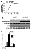
Fig. 3. Exogenous FRNK expression abrogates increased FAK activation in IPF lung fibroblasts
(A): HA-tagged FRNK expression was mediated by adenoviral vector (Ad-FRNK), and confirmed by immunofluorescence staining in normal human (NHL) and IPF (IPF) lung fibroblasts at the indicated multiplicity of infection (MOI). Digital images was obtained (10X), and plotted against the MOI (mean ± SE). (B): NHL (n = 2) and IPF (n = 2) lung fibroblasts were infected with either Ad-FRNK or control vector encoding the green fluorescent protein (Ad-GFP) at 100 MOI, and lysed after 36 hours. Equivalent amount of whole cell detergent lysates were Western blotted with the indicated antibodies. (C): Densitometry of FAK activation (normalized to total FAK protein) from Panel B. The data are represented as the fold activation of FAK relative to NHL group infected with control Ad-GFP.
Fig. 4
Fig. 4. Exogenous FRNK expression mediated by adenoviral vector (Ad-FRNK) abrogates increased cell migration in IPF lung fibroblasts, and inhibits PDGF-BB-stimulated cell migration in both normal human and IPF lung fibroblasts
Normal human lung fibroblasts (NHL, n = 5) and IPF lung fibroblasts (IPF, n = 9) were infected with Ad-FRNK or control Ad-GFP, and subjected to the monolayer wound healing assay as shown in Fig. 1. (A): wounded NHLs and (B): wounded IPFs were treated without or with PDGF-BB (4 ng/ml) for 24 hours in serum-free media (SFM) with 1 % BSA at 37°C. The wound area was monitored and quantified as in Fig. 1. Data were pooled and plotted (mean ± SE) as the % of wound area covered over 24 hours relative to NHL in basal (SFM) condition. * and # represent p < 0.001.
Fig. 5
Fig. 5. Exogenous FRNK expression mediated by adenoviral vector (Ad-FRNK) inhibits cell migration toward fibronectin and collagen in both normal human and IPF lung fibroblasts
Normal human (NHL, n = 5) and IPF (IPF, n = 9) lung fibroblasts were infected with Ad-FRNK or control Ad-GFP as in Fig. 4. Cells were then plated onto filters coated on the bottom surface with 10 µg/ml of fibronectin (shown in Panel A:) or 10 µg/ml of collagen (shown in Panel B:), allowed to migrate for 6 hours (37°C, 5% CO2), and haptotactic cell migration was determined as described in the “METHODS”. The data were analyzed and presented as the mean ± SE. * represents p < 0.01.
Fig. 6
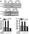
Fig. 6. Increased Rac and Rho activation were found in IPF lung fibroblasts, and exogenous FRNK expression abrogates increased Rac and Rho activation in IPF lung fibroblasts
(A): Normal human (NHL, n = 5) and IPF (IPF, n = 5) lung fibroblasts were lysed at 36 hours after Ad-FRNK or control Ad-GFP infection, as in Fig. 3. Rac and Rho activation were examined as described in the “METHODS”. Equivalent amount of whole cell detergent lysates were also Western blotted for total Rac and Rho. NHL (n=5) and IPF (n=5) lung fibroblasts without adenoviral construct infection (No Ad) served as a control, and representative image from one cell line each group was shown. (B) and (C): Densitometry of Rac and Rho activation from Panel A (normalized to total Rac and Rho protein, respectively). The data are represented as the fold change relative to the normal human lung fibroblasts infected with Ad-GFP.

Similar articles

Cited by

References

    1. King TE, Jr, Schwarz MI, Brown K, Tooze JA, Colby TV, Waldron JA, Jr, Flint A, Thurlbeck W, Chemiack RM. Idiopathic pulmonary fibrosis: relationship between histopathologic features and mortality. American Journal of Respiratory and Critical Care Medicine. 2001;164:1025–1032. - PubMed
    1. White ES, Thannickal VJ, Carskadon SL, Markwart S, Toews GB, Arenbert DA. Integrin alpha4beta1 regulates migration across basement membranes by lung fibroblasts: a role for phosphatase and tensin homologue deleted on chromosome 10. American Journal of Respiratory and Critical Care Medicine. 2003;168:436–442. - PMC - PubMed
    1. Parsons JT. Focal adhesion kinase: the first ten years. Journal of Cell Science. 2003;116:1409–1416. - PubMed
    1. Chen HC, Appeddu PA, Isoda H, Guan JL. Phosphorylation of tyrosine 397 in focal adhesion kinase is required for binding phosphatidylinositol 3-kinase. Journal of Biological Chemistry. 1996;271:26329–26334. - PubMed
    1. Ding Q, Grammer JR, Nelson MA, Guan JL, Stewart JE, Jr, Gladson CL. p27(Kip1) and cyclin D1 are necessary for focal adhesion kinase (FAK) regulation of cell cycle progression in glioblastoma cells propagated in vitro and in vivo in the scid mouse brain. Journal of Biological Chemistry. 2005;280:6802–6805. - PubMed

Publication types

MeSH terms

Substances