

Transgenic BDNF induces nerve fiber regrowth into the auditory epithelium in deaf cochleae
- PMID: 20109446
- PMCID: PMC2864331
- DOI: 10.1016/j.expneurol.2010.01.011
Transgenic BDNF induces nerve fiber regrowth into the auditory epithelium in deaf cochleae
Abstract
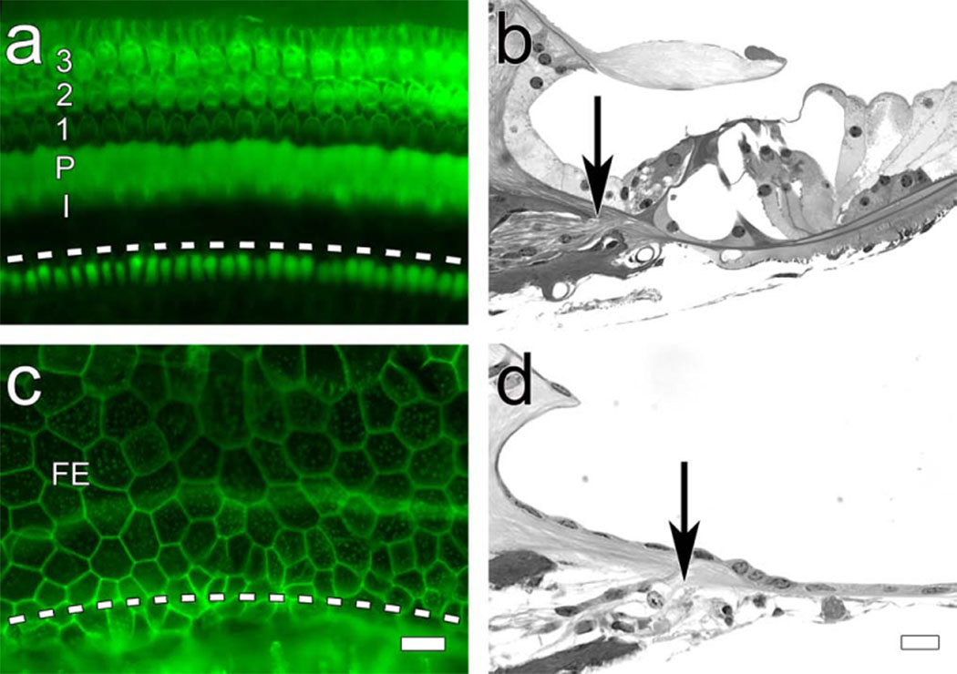
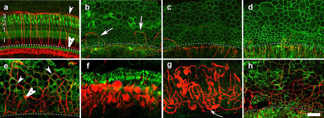
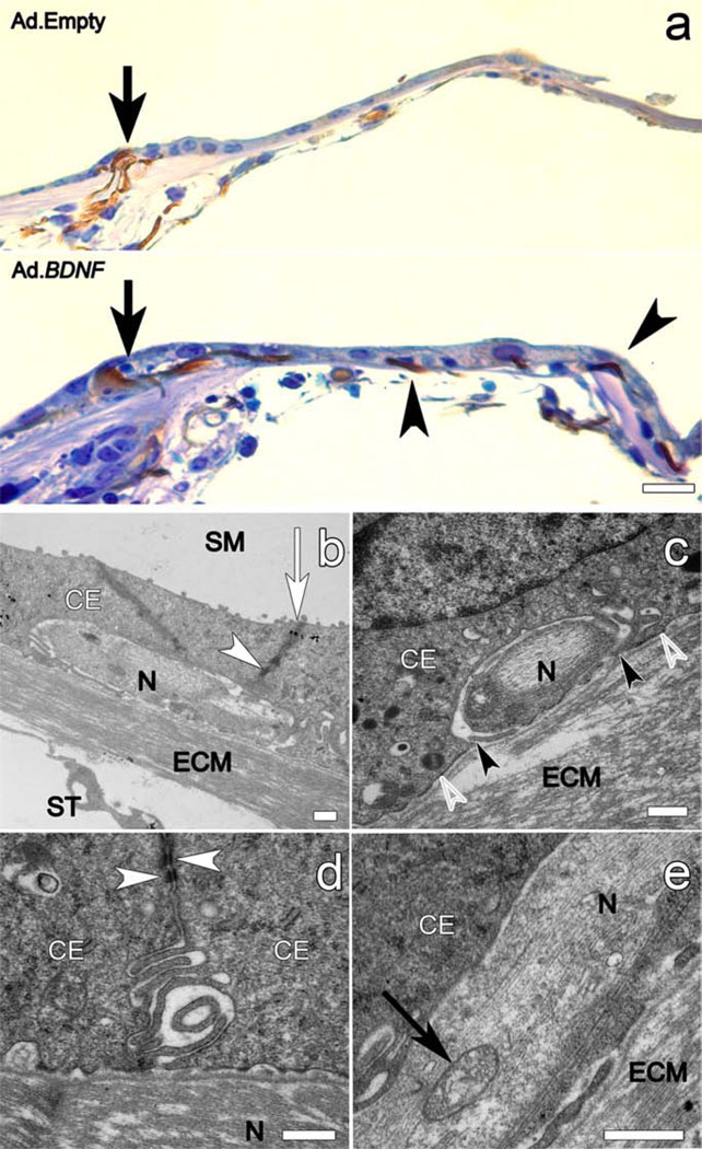
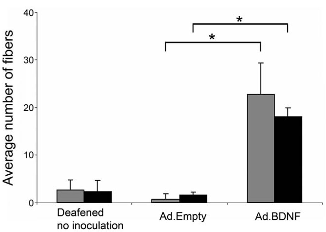
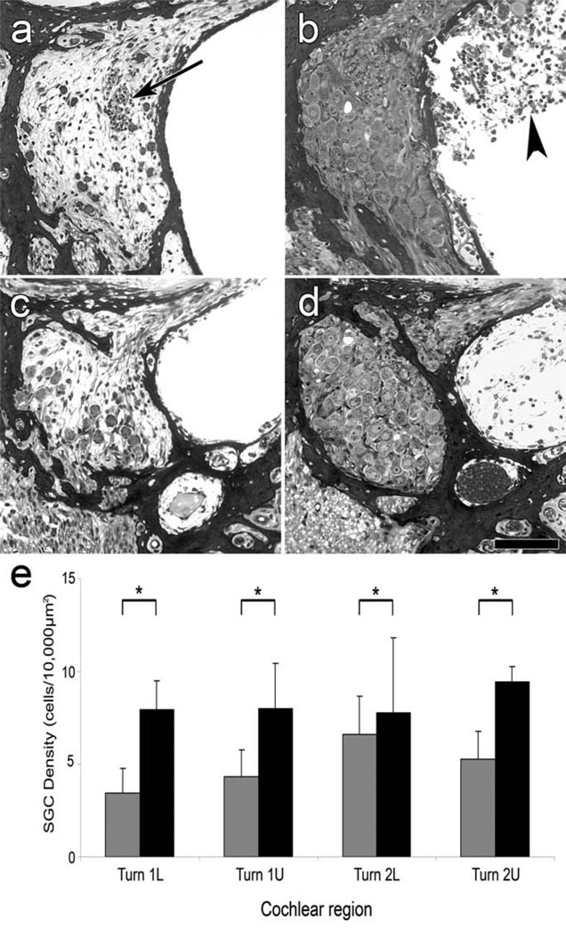
Sensory organs typically use receptor cells and afferent neurons to transduce environmental signals and transmit them to the CNS. When sensory cells are lost, nerves often regress from the sensory area. Therapeutic and regenerative approaches would benefit from the presence of nerve fibers in the tissue. In the hearing system, retraction of afferent innervation may accompany the degeneration of auditory hair cells that is associated with permanent hearing loss. The only therapy currently available for cases with severe or complete loss of hair cells is the cochlear implant auditory prosthesis. To enhance the therapeutic benefits of a cochlear implant, it is necessary to attract nerve fibers back into the cochlear epithelium. Here we show that forced expression of the neurotrophin gene BDNF in epithelial or mesothelial cells that remain in the deaf ear induces robust regrowth of nerve fibers towards the cells that secrete the neurotrophin, and results in re-innervation of the sensory area. The process of neurotrophin-induced neuronal regeneration is accompanied by significant preservation of the spiral ganglion cells. The ability to regrow nerve fibers into the basilar membrane area and protect the auditory nerve will enhance performance of cochlear implants and augment future cell replacement therapies such as stem cell implantation or induced transdifferentiation. This model also provides a general experimental stage for drawing nerve fibers into a tissue devoid of neurons, and studying the interaction between the nerve fibers and the tissue.
Copyright (c) 2009 Elsevier Inc. All rights reserved.
Figures

Similar articles
-
BDNF gene therapy induces auditory nerve survival and fiber sprouting in deaf Pou4f3 mutant mice.Sci Rep. 2012;2:838. doi: 10.1038/srep00838. Epub 2012 Nov 12. Sci Rep. 2012. PMID: 23150788 Free PMC article.
-
The effect of deafness duration on neurotrophin gene therapy for spiral ganglion neuron protection.Hear Res. 2011 Aug;278(1-2):69-76. doi: 10.1016/j.heares.2011.04.010. Epub 2011 May 1. Hear Res. 2011. PMID: 21557994 Free PMC article.
-
AAV-Mediated Neurotrophin Gene Therapy Promotes Improved Survival of Cochlear Spiral Ganglion Neurons in Neonatally Deafened Cats: Comparison of AAV2-hBDNF and AAV5-hGDNF.J Assoc Res Otolaryngol. 2019 Aug;20(4):341-361. doi: 10.1007/s10162-019-00723-5. Epub 2019 Jun 20. J Assoc Res Otolaryngol. 2019. PMID: 31222416 Free PMC article.
-
The Cochlear Spiral Ganglion Neurons: The Auditory Portion of the VIII Nerve.Anat Rec (Hoboken). 2019 Mar;302(3):463-471. doi: 10.1002/ar.23815. Epub 2018 May 4. Anat Rec (Hoboken). 2019. PMID: 29659185 Review.
-
Strategies to preserve or regenerate spiral ganglion neurons.Curr Opin Otolaryngol Head Neck Surg. 2005 Oct;13(5):294-300. doi: 10.1097/01.moo.0000180919.68812.b9. Curr Opin Otolaryngol Head Neck Surg. 2005. PMID: 16160524 Review.
Cited by
-
Research progress on flat epithelium of the inner ear.Physiol Res. 2020 Nov 16;69(5):775-785. doi: 10.33549/physiolres.934447. Epub 2020 Sep 9. Physiol Res. 2020. PMID: 32901490 Free PMC article. Review.
-
Expression of Oligodendrocyte Marker during Peripheral-Central Transitional Zone Formation of the Postnatal Mouse Cochlear Nerve.Otolaryngol Head Neck Surg. 2017 Sep;157(3):488-492. doi: 10.1177/0194599817718806. Epub 2017 Jul 11. Otolaryngol Head Neck Surg. 2017. PMID: 28695768 Free PMC article.
-
Conditional deletion of Atoh1 using Pax2-Cre results in viable mice without differentiated cochlear hair cells that have lost most of the organ of Corti.Hear Res. 2011 May;275(1-2):66-80. doi: 10.1016/j.heares.2010.12.002. Epub 2010 Dec 10. Hear Res. 2011. PMID: 21146598 Free PMC article.
-
Lithium alters the morphology of neurites regenerating from cultured adult spiral ganglion neurons.Hear Res. 2013 Oct;304:137-44. doi: 10.1016/j.heares.2013.07.001. Epub 2013 Jul 12. Hear Res. 2013. PMID: 23856237 Free PMC article.
-
A novel Atoh1 "self-terminating" mouse model reveals the necessity of proper Atoh1 level and duration for hair cell differentiation and viability.PLoS One. 2012;7(1):e30358. doi: 10.1371/journal.pone.0030358. Epub 2012 Jan 18. PLoS One. 2012. PMID: 22279587 Free PMC article.
References
-
- Aarnisalo AA, Pirvola U, Liang XQ, Miller J, Ylikoski J. Apoptosis in auditory brainstem neurons after a severe noise trauma of the organ of Corti: intracochlear GDNF treatment reduces the number of apoptotic cells. ORL J Otorhinolaryngol Relat Spec. 2000;62:330–334. - PubMed
-
- Altschuler RA, Cho Y, Ylikoski J, Pirvola U, Magal E, Miller JM. Rescue and regrowth of sensory nerves following deafferentation by neurotrophic factors. Ann N Y Acad Sci. 1999;884:305–311. - PubMed
-
- Altschuler RA, Cho Y, Ylikoski J, Pirvola U, Magal E, Miller JM. Rescue and regrowth of sensory nerves following deafferentation by neurotrophic factors. Ann N Y Acad Sci. 1999;884:305–311. - PubMed
-
- Beyer LA, Odeh H, Probst FJ, Lambert EH, Dolan DF, Camper SA, Kohrman DC, Raphael Y. Hair cells in the inner ear of the pirouette and shaker 2 mutant mice. J Neurocytol. 2000;29:227–240. - PubMed
-
- Bichler E, Spoendlin H, Rauchegger H. Degeneration of cochlear neurons after amikacin intoxication in the rat. Archives of Oto-Rhino-Laryngology. 1983;237:201–208. - PubMed
Publication types
MeSH terms
Substances
Grants and funding
LinkOut - more resources
Full Text Sources
Other Literature Sources
Medical
Molecular Biology Databases
