. 2010 May;149(2):152-61.
doi: 10.1016/j.virusres.2010.01.011. Epub 2010 Jan 28.

Genomic characterization of human adenovirus 36, a putative obesity agent

Affiliations

Genomic characterization of human adenovirus 36, a putative obesity agent

John Arnold et al. Virus Res. 2010 May.

Abstract

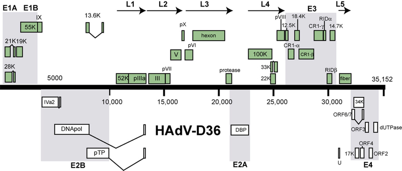
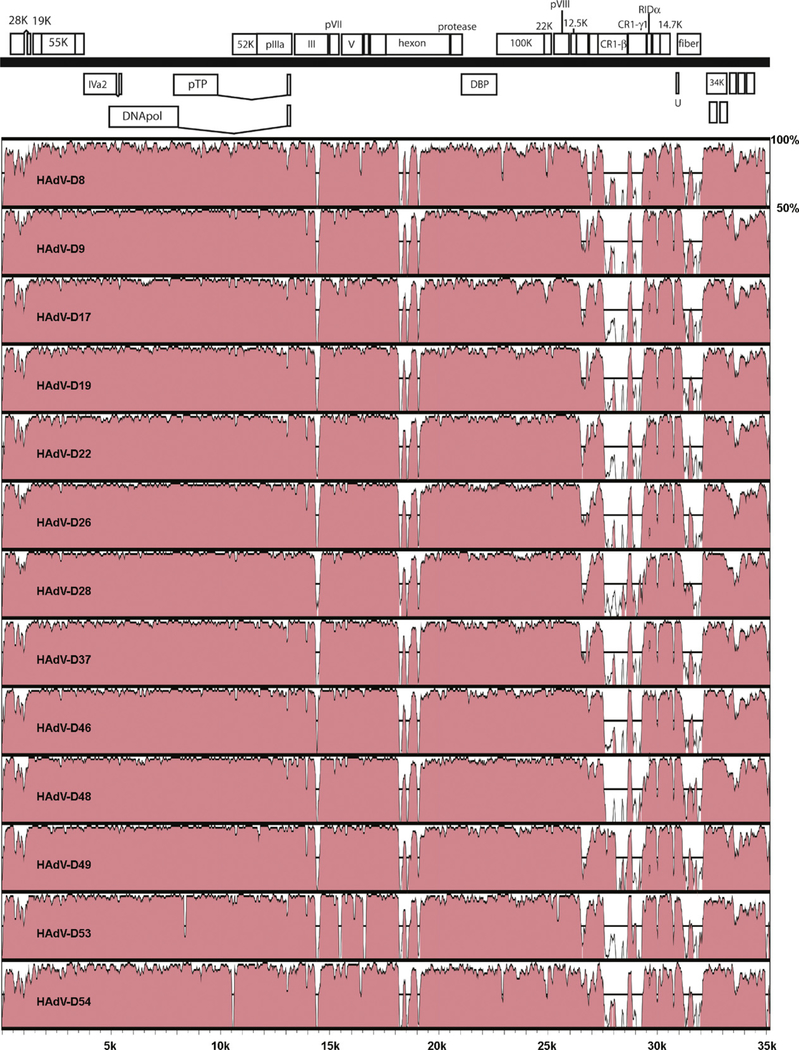
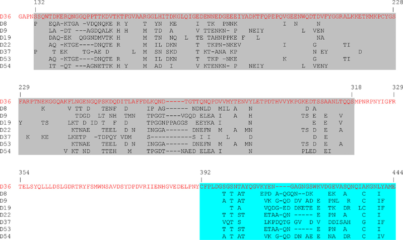
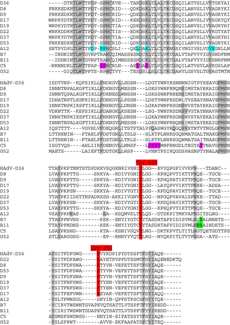
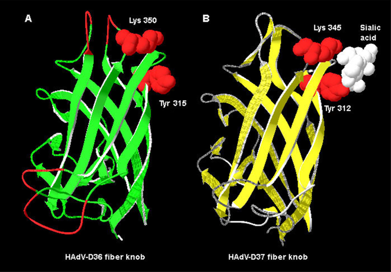
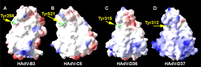
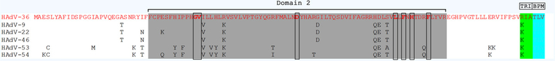
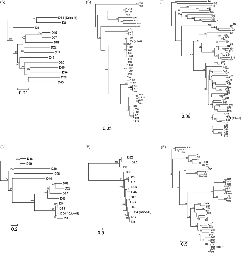
Increased levels of serum antibody titers against human adenovirus 36 (HAdV-D36) are associated with human obesity and experimental obesity in laboratory animals. While HAdV-D36 has been studied as an infectious agent implicated in obesity for over a decade, the complete genome sequence and its analysis have yet to be reported. A detailed analysis of the genome sequence of HAdV-D36 may be important to understand its role in obesity. Genomic and bioinformatic comparisons with other HAdVs identified differences that suggested unique functions. Global pairwise genome alignment with all sequenced human adenovirus D (HAdV-D) genomes revealed areas of nonconserved sequences in the hexon, E3 CR1 beta, E3 CR1 gamma, and fiber genes. Phylogenetic analysis of all HAdV-D36 proteins confirmed that this virus belongs to species Human adenovirus D. This genomic analysis of HAdV-D36 provides an important tool for comprehending the role that this unique adenovirus may play in human obesity. Low amino acid sequence identity in the E3 CR1 beta and CR1 gamma genes may suggest distinctive roles for these proteins. Furthermore, the predicted molecular models of the HAdV-D36 fiber protein seem to implicate a unique tissue tropism for HAdV-D36.


Figures

Fig. 1.
Fig. 1.
Map of predicted open reading frames and their identities in the genome of HAdV-D36.
Fig. 2.
Fig. 2.
Pairwise comparison of adenovirus genomes from species HAdV-D to HAdV-D36.
Fig. 3.
Fig. 3.
Multiple-sequence alignment of hexon loops 1 and 2 from species HAdV-D. Loop 1 is highlighted in grey and loop 2 is highlighted in aqua. Parts of the conserved regions were deleted to save space. (For interpretation of the references to color in this figure legend, the reader is referred to the web version of the article.)
Fig. 4.
Fig. 4.
Multiple alignment of adenovirus fiber knob sequences from species HAdV-D. Alignment contains adenovirus fiber knob from sequences from species HAdV-D (−8, −9, −17, −19, −22, −37, and −53), HAdV-A, HAdV-B, HAdV-C, and HAdV-G. Tyr315 and Lys350, the vital amino acids that form the sialic acid binding site, are highlighted in red. The conserved residues are highlighted in grey. Amino acids contacting CAR in the HAdV-A12–CAR complex crystal structure are shown on an aqua background. The residues in the HAdV-C5 fiber protein that were found to interact with CAR by mutagenesis (Kirby et al., 1999, 2000) are highlighted in magenta. Residues that interact with CD46 are highlighted in green. (For interpretation of the references to color in this figure legend, the reader is referred to the web version of the article.)
Fig. 5.
Fig. 5.
Molecular model of the HAdV-D36 fiber knob. (A) Molecular model of HAdV-D36 knob with Tyr315 and Lys350 highlighted in red. (B) Structure of the HAdV-D37 fiber knob bound to sialic acid (white). Lys345 and Tyr312, which bind sialic acid are highlighted in red. (For interpretation of the references to color in this figure legend, the reader is referred to the web version of the article.)
Fig. 6.
Fig. 6.
Electrostatic surface potential of HAdV-D36 fiber knob. Side view of fiber knobs from (A) HAdV-B3, (B) HAdV-C5, (C) HAdV-D36, and (D) HAdV-D37. The figure was prepared with DeepView (http://www.expasy.org/spdbv/) (Guex and Peitsch, 1997). The green dots demarcate the location of the conserved Tyr residues which are associated with binding sialic acid. Coloring of the surface potential ranges from +5 kT (blue) to −5 kT (red). (For interpretation of the references to color in this figure legend, the reader is referred to the web version of the article.)
Fig. 7.
Fig. 7.
Multiple-sequence alignment of adenovirus dUTPase (E4-ORF1) proteins from species HAdV-D. Domain 2 is highlighted in grey, TRI is highlighted in green, and the PBM element is highlighted in aqua. Amino acids that are critical for the function of domain 2 (G40, V41, D65, L89, F91, H93, and F97) are in bold and in black boxes. (For interpretation of the references to color in this figure legend, the reader is referred to the web version of the article.)
Fig. 8.
Fig. 8.
Phylogenetic analysis of HAdV-D36. Analysis of HAdV-D36 is based on the nucleic acid sequence of (A) complete genomes, as well as the predicted amino acid sequences of (B) DNA polymerase, (C) hexon, (D) CR1β, (E) CR1γ, and (F) fiber.

Similar articles

Cited by

References

    1. Atkinson RL, Dhurandhar NV, Allison DB, Bowen RL, Israel BA, Albu JB, Augustus AS, 2005. Human adenovirus-36 is associated with increased body weight and paradoxical reduction of serum lipids. Int. J. Obes. (Lond.) 29 (3), 281–286. - PubMed
    1. Bouchard C, 2009. Childhood obesity: are genetic differences involved? Am. J. Clin. Nutr 89 (5), 1494S–1501S. - PMC - PubMed
    1. Brown WV, Fujioka K, Wilson PW, Woodworth KA, 2009. Obesity: why be concerned? Am. J. Med 122 (4 (Suppl. 1)), S4–S11. - PubMed
    1. Brudno M, Do CB, Cooper GM, Kim MF, Davydov E, Green ED, Sidow A, Batzoglou S, 2003. LAGAN and Multi-LAGAN: efficient tools for large-scale multiple alignment of genomic DNA. Genome Res. 13 (4), 721–731. - PMC - PubMed
    1. Chung SH, Frese KK, Weiss RS, Prasad BV, Javier RT, 2007. A new crucial protein interaction element that targets the adenovirus E4-ORF1 oncoprotein to membrane vesicles. J. Virol 81 (9), 4787–4797. - PMC - PubMed

Publication types

Associated data

LinkOut - more resources