Review
. 2010 Apr;90(4):471-97.
doi: 10.1016/j.pneurobio.2010.01.001. Epub 2010 Jan 29.

The functional organisation of glia in the adult brain of Drosophila and other insects

Affiliations
Review

The functional organisation of glia in the adult brain of Drosophila and other insects

Tara N Edwards et al. Prog Neurobiol. 2010 Apr.

Abstract

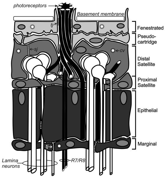
This review annotates and categorises the glia of adult Drosophila and other model insects and analyses the developmental origins of these in the Drosophila optic lobe. The functions of glia in the adult vary depending upon their sub-type and location in the brain. The task of annotating glia is essentially complete only for the glia of the fly's lamina, which comprise: two types of surface glia-the pseudocartridge and fenestrated glia; two types of cortex glia-the distal and proximal satellite glia; and two types of neuropile glia-the epithelial and marginal glia. We advocate that the term subretinal glia, as used to refer to both pseudocartridge and fenestrated glia, be abandoned. Other neuropiles contain similar glial subtypes, but other than the antennal lobes these have not been described in detail. Surface glia form the blood brain barrier, regulating the flow of substances into and out of the nervous system, both for the brain as a whole and the optic neuropiles in particular. Cortex glia provide a second level of barrier, wrapping axon fascicles and isolating neuronal cell bodies both from neighbouring brain regions and from their underlying neuropiles. Neuropile glia can be generated in the adult and a subtype, ensheathing glia, are responsible for cleaning up cellular debris during Wallerian degeneration. Both the neuropile ensheathing and astrocyte-like glia may be involved in clearing neurotransmitters from the extracellular space, thus modifying the levels of histamine, glutamate and possibly dopamine at the synapse to ultimately affect behaviour.


Figures

Fig. 1
Fig. 1. Glia in the adult fly lamina
The lamina of the adult optic lobe of the fly is populated by six distinct classs of glia. These include (from distal to proximal) two types of surface glia - the fenestrated and pseudocartridge glia; two types of cortex glia - the distal and proximal satellite glia; and two types of neuropile glia - the epithelial and marginal glia. Septate junctions (sj) connect the distal satellite glia and are an integral part of the blood brain barrier. Tight junctions (tj) are more common in the proximal glial layers. The outermost glia also contain clathrin coated vesicles (cv) and engage in clathrin-mediated endocytosis. Figure modified from Saint-Marie and Carlson (1993a).
Fig. 2
Fig. 2. Glia in the larval visual system
The larval visual system becomes populated by glia from at least two different sources, the eye disc and in the developing optic lobe, the glial precursor centre (GPC). A. The glia of the eye disc (brown nuclei) all originate in the optic stalk (osg) and migrate into the eye disc. There are four types of differentiated glia in the eye disc: two large basally-located carpet glia (cg, outline in dashed lines), the eye disc marginal glia (edmg; not to be confused with the lamina marginal glia), wrapping glia (wg) and surface glia (surg). Undifferentiated glia (udg, outline in dashed lines) migrate along the basal surface of the eye disc, below the carpet glia, until they come into contact with newly differentiated photoreceptors (R) just posterior to the morphogenetic furrow (mf). Differentiating glia then migrate apically and develop extensions to surround photoreceptor axons, becoming wrapping glia. A grey dashed line indicates a rotation of the brain relative to the optic lobe for illustration purposes, but both wrapping and carpet glia extend into the optic lobes. In the optic lobe, glia are derived from the GPC which lies proximal to the lamina furrow (LF) and the Outer Optic Anlage (OOA), from whence neuronal precursors arise. Three types of lamina glia (dark blue) derive from the GPC; these include at least some of the satellite glia (sg), as well as the epithelial (eg) and marginal glia (mg). The GPC also gives rise to the glia lining the medulla neuropile (light blue) including the outer chiasmal glia (xg) and the medulla neuropile glia (mng). Cells underlying the larval marginal glia are usually labelled medulla glia in the literature, failing to acknowledge that in the adult an additional layer of glia, those of the outer chiasm ‘small’ and ‘giant’ glia (Tix et al., 1997), lies between the marginal glia and medulla glia. Lamina precursor cells (LPC) are displaced to the lamina where, as lamina neurons (ln), their cell bodies come to lie between the satellite and the epithelial glia. Likewise, medulla precusor cells (MPC), possibly ganglion mother cells, ultimately give rise to medulla neurons (mn). Subperineurial glia (supng; pink nuclei) derived from the epithelium and mesodermally derived perineurial glia (png; grey nuclei) surround the entire optic lobe as a sheath to form distinct components of the blood brain barrier. The inner glia of the optic stalk and the medulla cortex glia are not illustrated. B. A cross section of the eye disc shows the relative apical/basal locations of the glia and their locations in relation to the photoreceptors. Figures modified from originals in Chotard et al. (2005) and Silies et al. (2007).
Fig. 3
Fig. 3. The glial anatomy of Drosophila
There are three classes of glia in the insect brain, surface, cortex and neuropile. Figures A and B depict a frontal view of (A) the Drosophila head and (B) underlying brain neuropiles. The lamina (La), medulla (Me), lobula (Lo) and lobula plate (LoP) constitute the visual protocerebrum (B and C) which underlie the retina (Re). The antennal lobes (AL) and mushroom bodies (MB) constitute the deutocerebrum, while all other neuropiles (not illustrated here) including the subesophogeal ganglion (SoG) belong to the tritrocerebrum. A horizontal section through the visual neuropiles (C, section plane C in B) shows the nuclear location of surface (light grey), chiasm (light grey), cortex (medium grey) and neuropile (dark grey) glia relative to their respective neuropiles and their associated neurons (nuclei in white). Subperineurial surface glia and cortex glia (called satellite glia when associated with the visual system) are sparse and only a few satellite glia are required to surround many neuron cell bodies in the cortex. Chiasm glia lie in two locations: between the lamina and medulla neuropile (first optic chiasm) and the medulla and lobula/lobula plate neuropiles (second optic chiasm), in both cases forming an anterior to posterior glial boundary. Within the first optic chiasm two types of glia can be distinguished, small and giant. Neuropile glia lie amongst the axon terminals but for some optic lobe neuropiles no distinction has yet been made between their ensheathing and astrocyte-like glia. Chandelier glia, which have been detected in the neuropile of other Diptera, but have not yet been described in Drosophila, are illustrated in dotted outline at the base of the medulla. On the other hand, within the antennal lobes (D) both types of neuropile glia (dark grey nuclei): ensheathing (en) glia, which wrap the neuropile, and astrocyte-like glia (ag), which extend processes amongst the glomeruli, can be distinguished using different GAL4 driver lines. Outside the neuropile, neuronal cell bodies (white) are ensconced in extensions from cortex glia (ctx; medium grey nuclei) which separate the antennal lobe from neighbouring neuropiles and the oesophagus (eso). Nerve layer glia lie at the base of the antennal nerve (AN) where the nerve enters the antennal lobe, while surface glia (sg) ensheathe the nerve, and tract glia (tg) lie at the edge of the antennal lobe commissures.
Fig. 4
Fig. 4. Visual system function is dependent upon an intimate association of photoreceptors and glia
A. Histamine (HA) is synthesized from histidine (His) in the photoreceptor by the enzyme histadine decarboxylase (HDC). HA is released from vesicles at the photoreceptor T-bar ribbon. In the synaptic cleft it can act on HA gated chloride channels at the surface of the monopolar cells (L1,L2) or the glia. Excess HA is taken up by the glia where it is then inactivated by conjugation to β-alanine (β-ala) by the protein Ebony. β-ala is produced in the glia from decarboxylation of aspartate (Asp) by Black, or by catabolism of uracil (Ura) along a dihydropyrimidine dehydrogenase (DPD+) dependent pathway (Rawls, 2006). The HA- β-alanyl conjugate, called carcinine (CA), is shuttled from the glia back into the photoreceptor by an unknown mechanism (path 1) or possibly via the capitate projection (cp, path 2), where clathrin-mediated endocytosis of coated vesicles (cv) takes place. In the photoreceptor HA is liberated from CA by Tan. Liberated HA can then be pumped back into recycled vesicles and prepared once again for release at the synapse. B. An EM cross-section of the Drosophila lamina. Photoreceptors (blue) synapse (arrow) onto paired monopolar neurons (L, magenta). Epithelial glia (green) surround the cartridge and invests areas close to synapses. Glia invaginate into photoreceptors at specialised sites called capitate projections (double arrowheads). C. The inverted extracellular response (ERG) recorded from the eye of Drosophila is triggered by a light flash (*) and consists of the combined negative sustained response (sp) of the photoreceptors and the hyperpolarizing (“on”) and depolarizing (“off”) responses of the lamina. The “on” response is modulated by activation of HisCl1 receptors on the epithelial glia.

Similar articles

Cited by

References

    1. Andretic R, Hirsh J. Circadian modulation of dopamine receptor responsiveness in Drosophila melanogaster. Proc. Natl. Acad. Sci. U.S.A. 2000;97:1873–1878. - PMC - PubMed
    1. Artalejo CR, Henley JR, McNiven MA, Palfrey HC. Rapid endocytosis coupled to exocytosis in adrenal chromaffin cells involves Ca2+, GTP, and dynamin but not clathrin. Proc Natl Acad Sci U S A. 1995;92:8328–8332. - PMC - PubMed
    1. Augustin H, Grosjean Y, Chen K, Sheng Q, Featherstone DE. Nonvesicular release of glutamate by glial xCT transporters suppresses glutamate receptor clustering in vivo. J. Neurosci. 2007;27:111–123. - PMC - PubMed
    1. Awasaki T, Tatsumi R, Takahashi K, Arai K, Nakanishi Y, Ueda R, Ito K. Essential role of the apoptotic cell engulfment genes draper and ced-6 in programmed axon pruning during Drosophila metamorphosis. Neuron. 2006;50:855–967. - PubMed
    1. Awasaki T, Lai S-L, Ito K, Lee T. Organization and postembryonic development of glial cells in the adult central brain of Drosophila. J. Neurosci. 2008;28:13742–13753. - PMC - PubMed

Publication types

Substances

LinkOut - more resources