Review
. 2010 May;21(3):276-82.
doi: 10.1016/j.semcdb.2010.01.016. Epub 2010 Jan 28.

Kinesin-13s in mitosis: Key players in the spatial and temporal organization of spindle microtubules

Affiliations
Review

Kinesin-13s in mitosis: Key players in the spatial and temporal organization of spindle microtubules

Stephanie C Ems-McClung et al. Semin Cell Dev Biol. 2010 May.

Abstract

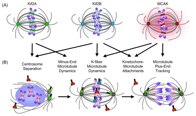
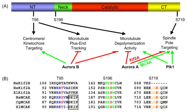
Dynamic microtubules are essential for the process of mitosis. Thus, elucidating when, where, and how microtubule dynamics are regulated is key to understanding this process. One important class of proteins that directly regulates microtubule dynamics is the Kinesin-13 family. Kinesin-13 proteins induce depolymerization uniquely from both ends of the microtubule. This activity coincides with their cellular localization and with their ability to regulate microtubule dynamics to control spindle assembly and kinetochore-microtubule attachments. In this review, we highlight recent findings that dissect the important actions of Kinesin-13 family members and summarize important studies on the regulation of their activity by phosphorylation and by protein-protein interactions.


Figures

Fig. 1
Fig. 1
Localization and function of Kinesin-13s in mitosis. (A) The vertebrate Kinesin-13s have overlapping localizations at spindle poles and kinetochores/centromeres. MCAK is also found in the cytoplasm and at the plus-ends of microtubules. Kif2A localizations are shown in green, Kif2B in teal, and MCAK in red. Microtubules are shown in gray and chromosomes are shown in blue. (B) Summary of vertebrate Kinesin-13 function during mitosis. Spindle structures and Kinesin-13 models are colored as in (A) with kinetochores depicted in pink. Models of the Kinesin-13s are shown at their predominant site(s) of function in prophase, metaphase, and anaphase.
Fig. 2
Fig. 2
Kinesin-13 localization and function is regulated by conserved phosphorylation sites. (A) Schematic of secondary amino acid structure of Kinesin-13s and summary of Aurora B, Aurora A, and Plk1 phosphorylation effects. NT, N-terminal domain; and CT, C-terminal domain. Only the Xenopus MCAK phosphorylation sites are depicted for clarity. Green arrows depict promotion and red lines depict inhibition of localization or activity. (B) The T95, S196, and S719 MCAK phosphorylation sites are highly conserved in Kif2A, whereas only the S196 site is conserved in Kif2B. For HsKif2A, S157 is equivalent to S132 as reported in Knowlton, et al. [70]. Phosphorylation sites are colored red, and the surrounding conserved residues are colored blue for the T95 site, green for the S196 site, and orange for the S719 site. The SxIP microtubule tip localization signal is boxed, and is only conserved in MCAK. Hs, human; Xl, frog (Xenopus laevis); and Cg, Chinese hamster. Genbank accession numbers used for alignment: HsKif2B, NM032559; HsKif2A, NM004520; XlKif2A, BC057698; HsMCAK, NM006845; CgMCAK, U11790; and XlMCAK, BC044976.

Similar articles

Cited by

References

    1. Mitchison TJ, Kirschner MW. Dynamic instability of microtubule growth. Nature. 1984;312:237–42. - PubMed
    1. Belmont LD, Hyman AA, Sawin KE, Mitchison TJ. Real-time visualization of cell cycle dependent changes in microtubule dynamics in cytoplasmic extracts. Cell. 1990;62:579–89. - PubMed
    1. Saxton WM, Stemple DL, Leslie RJ, Salmon ED, Zavortink M, McIntosh JR. Tubulin dynamics in cultured mammalian cells. J Cell Biol. 1984;99:2175–86. - PMC - PubMed
    1. Zhai Y, Kronebusch PJ, Simon PM, Borisy GG. Microtubule dynamics at the G2/M transition: Abrupt breakdown of cytoplasmic microtubules at nuclear envelope breakdown and implications for spindle morphogenesis. J Cell Biol. 1996;135:201–14. - PMC - PubMed
    1. Rusan NM, Fagerstrom CJ, Yvon AM, Wadsworth P. Cell cycle-dependent changes in microtubule dynamics in living cells expressing green fluorescent protein-alpha tubulin. Mol Biol Cell. 2001;12:971–80. - PMC - PubMed

Publication types

LinkOut - more resources