Review
. 2010 Jan;125(1):16-29.e1-11; quiz 30-1.
doi: 10.1016/j.jaci.2009.11.008.

An update on the genetics of atopic dermatitis: scratching the surface in 2009

Affiliations
Review

An update on the genetics of atopic dermatitis: scratching the surface in 2009

Kathleen C Barnes. J Allergy Clin Immunol. 2010 Jan.

Abstract

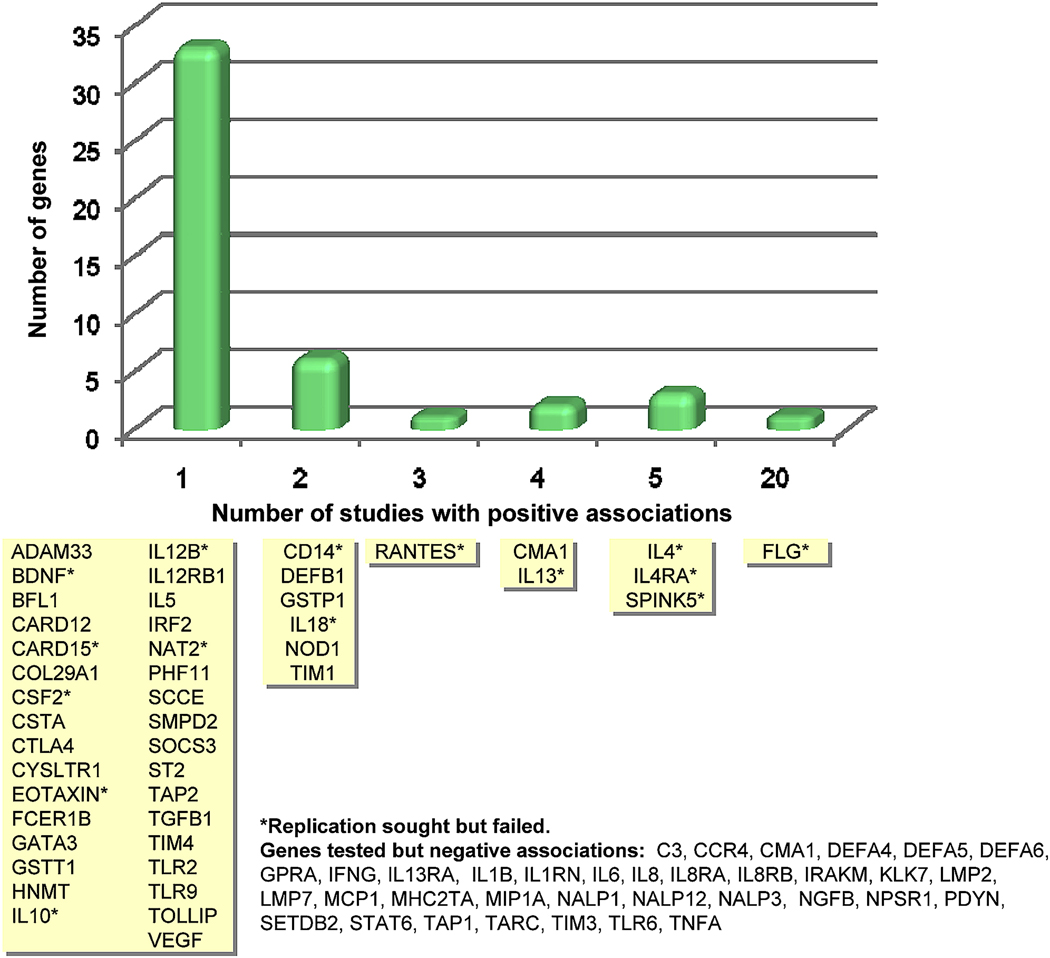
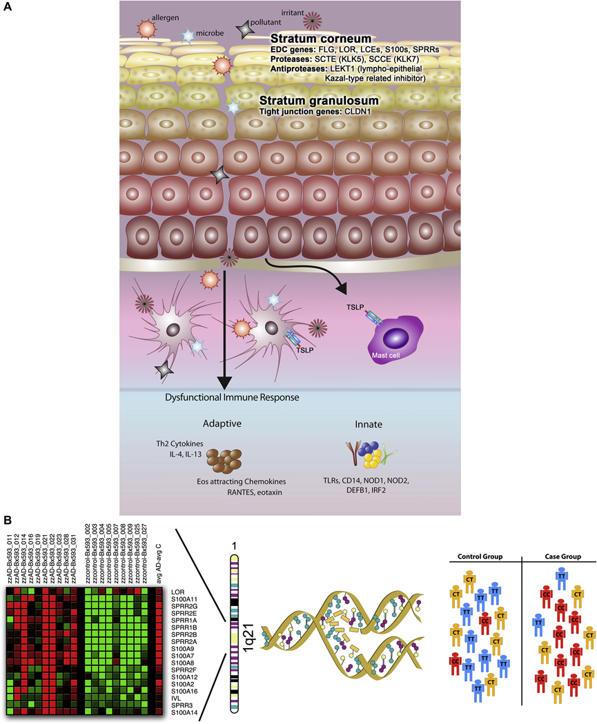
A genetic basis for atopic dermatitis (AD) has long been recognized. Historic documents allude to family history of disease as a risk factor. Before characterization of the human genome, heritability studies combined with family-based linkage studies supported the definition of AD as a complex trait in that interactions between genes and environmental factors and the interplay between multiple genes contribute to disease manifestation. A summary of more than 100 published reports on genetic association studies through mid-2009 implicates 81 genes, in 46 of which at least 1 positive association with AD has been demonstrated. Of these, the gene encoding filaggrin (FLG) has been most consistently replicated. Most candidate gene studies to date have focused on adaptive and innate immune response genes, but there is increasing interest in skin barrier dysfunction genes. This review examines the methods that have been used to identify susceptibility genes for AD and how the underlying pathology of this disease has been used to select candidate genes. Current challenges and the potential effect of new technologies are discussed.


Figures

Figure 1
Figure 1. Methods for detecting disease susceptibility genes
Panel A, genome-wide linkage method, relying on polymorphic microsatellites, referred to as ‘short tandem repeat polymorphisms’, or STRPs, evenly spaced across the chromosomes, typically including ~350 STRPs. Panel B, the candidate gene approach, whereby genetic markers, usually easily typed substitutions (i.e., single nucleotide polymorphisms, or SNPs) or structural variants (i.e., insertions/deletions), are selected within and flanking a candidate gene of interest. Panel C, genome-wide association studies, or GWAS, using hundreds of thousands and up to 1M SNPs to fully cover an individual’s genome, is a hypothesis-free, systematic search across the genome to identify novel associations with common diseases using a set of haplotype tagging SNPs (htSNPs), indicated as blue arrows. htSNPs represent a relatively small subset of SNPs of the potential 2.5 million SNPs in the public database (i.e., needed to uniquely identify a complete haplotype. The premise behind htSNPs is the observation that, when SNPs are in linkage disequilibrium (LD) with each other and form haplotypes, there is redundant information contained within the haplotype, because several of the SNPs will always occur together. Thus, a marker at one locus can reasonably predict a marker(s) that will occur at the linked locus nearby.
Figure 2
Figure 2. Summary of genome-wide linkage studies of atopic dermatitis (AD)
Representation of the 23 human chromosomes, highlighting those loci for which genome screens have identified linkage to AD. Loci are mapped to short or long chromosomal arms and color-coded according to the studies listed in the legend (Refs. –20).
Figure 3
Figure 3. Genes associated with atopic dermatitis in at least one published study
Genes are grouped according to how many positive association studies have been reported (see Table EI in this article’s Online Repository at www.jacionline.org for a complete summary of 111 published studies). The Y-axis indicates the number of genes (corresponding to the yellow boxes) for each time positive association was reported.
Figure 4
Figure 4. Networks revealed through the Ingenuity Pathways Analysis based on 81 AD genes established in genetic association studies
48 genes clustered into two major networks associated with immune dysregulation. Panel A, antigen presentation and cell-mediated and humoral immune response pathway. Significantly associated genes are highlighted in dark blue. Panel B, cell signaling and interaction, cellular movement, and hematological system development and function pathway. Significantly associated genes are highlighted in dark green. For both panels, genes or gene products are represented as nodes (shapes; see legend) that represent the functional class of the gene product. Relationship between genes is presented as solid lines with arrows (direct activation) and arrows point to the element on which an action is performed. All relationships (or lines) are supported by at least 1 reference (numbers of references in parentheses). Annotation of relationship (labels) between the nodes: A – activation, E - expression, I – inhibition, MB - group/complex membership, PD - Protein-DNA binding, PP - protein-protein binding.
Figure 5
Figure 5. Genetic epidemiological approaches toward identification of genes conferring AD susceptibility
In this summary of approaches used to date to identify candidate genes for AD, the first consideration is the complex pathology of AD (panel A), which includes defects and damage at the epithelial barrier, and penetration of allergens, microbes, pollutants and irritants into the epidermis and dermis, ultimately interacting with antigen presentation cells (i.e., Langerhans cells and dermal dendritic cells). The ‘brick wall-like’ structure of the stratum corneum is compromised, possibly by defects in genes residing in the epidermal differentiation complex (EDC), including FLG, LOR, LCEs, S100s, SPRRs, SCTE, and SCCE. Additional candidates include tight junctions (TJ), proteins that constitute the “gate” to the passage of water, ions and solutes through the paracellular pathway in the stratum granulosum (i.e., CLDN1). A defective innate immune response (involving, for example, the TLRs, CD14, NOD1, NOD2, DEFB1, and IRF2) may contribute to a heightened IgE-mediated, systemic Th2 response. Combining a candidate gene approach with robust, genomewide gene expression profiling has the potential of both elucidating novel candidates and validating suspected candidates, as illustrated in panel B, in which genes in the EDC on chromosome 1q21 are significantly differentially expressed in a human skin biopsies taken from AD patients compared to healthy nonatopic controls (courtesy L.A. Beck). Candidate genes are selected, and substitutions (i.e., single nucleotide polymorphisms, or SNPs) and simple structural variants (i.e., insertions/deletions, repeats) are genotyped in large populations selected for AD and tests for association are performed.

Similar articles

Cited by

References

    1. Smith WD. Hippocrates. Cambridge, MA: Harvard University Press; 1994.
    1. Besnier E. Premier note et observations preliminaire pour service d-introduction a letute des prurigos diathesiques. Ann Dermatol Syphil. 1892;3:634–638.
    1. Coca AF. Specific Diagnosis and Treatment of Allergic Diseases of the Skin. JAMA. 1934;103(17):1275–1277.
    1. Dold S, Wjst M, Mutius Ev, Reitmeir P, Stiepel E. Genetic risk for asthma, allergic rhinitis, and atopic dermatitis. Archives of Disease in Childhood. 1992;67:1018–1022. - PMC - PubMed
    1. Sneddon IB. The management of infantile eczema. Vol. 226. Medical Press; 1951. pp. 329–333. - PubMed

Publication types