

Magnetic resonance spectroscopy imaging of the newborn brain--a technical review
- PMID: 20109969
- PMCID: PMC2842012
- DOI: 10.1053/j.semperi.2009.10.003
Magnetic resonance spectroscopy imaging of the newborn brain--a technical review
Abstract
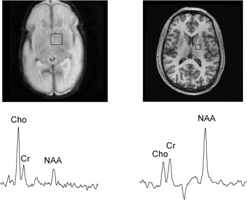
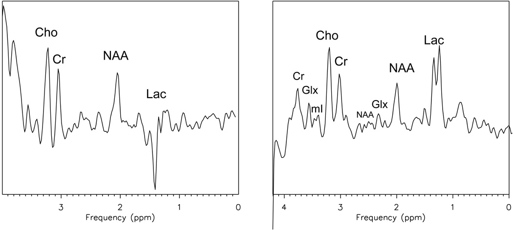
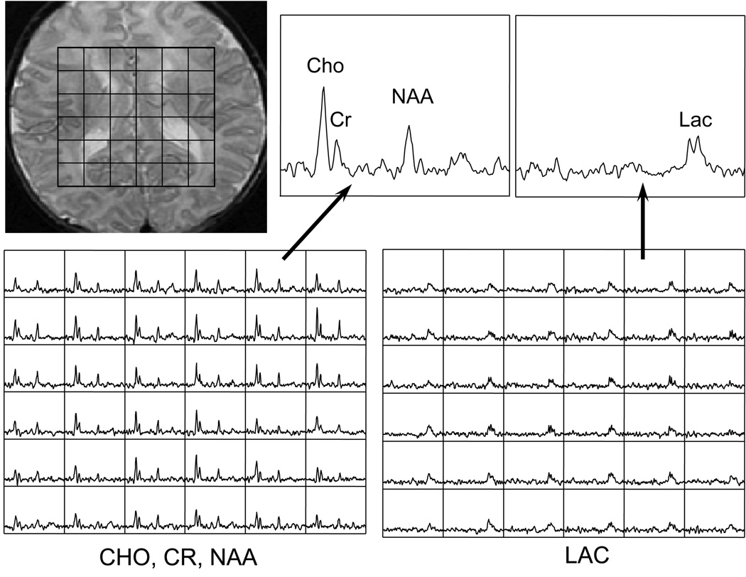
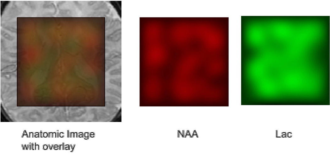
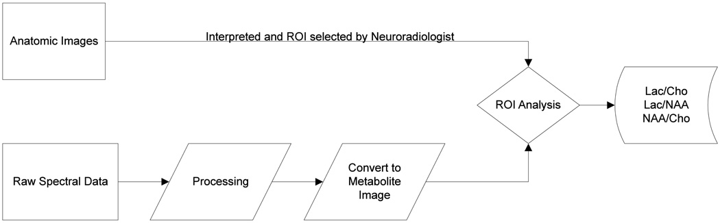
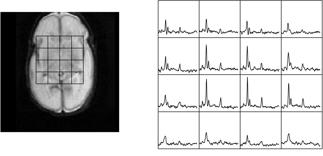
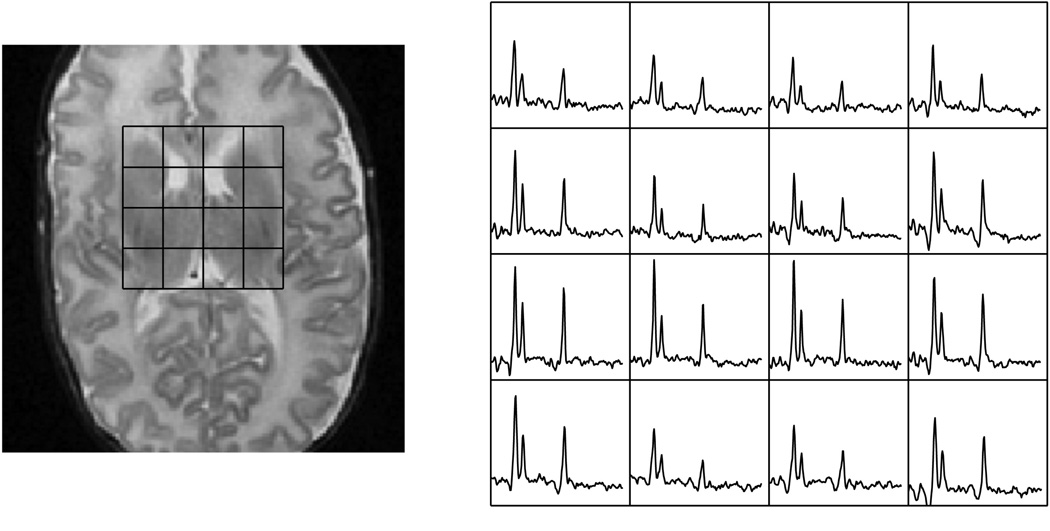
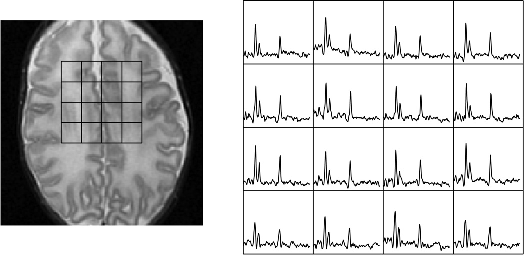
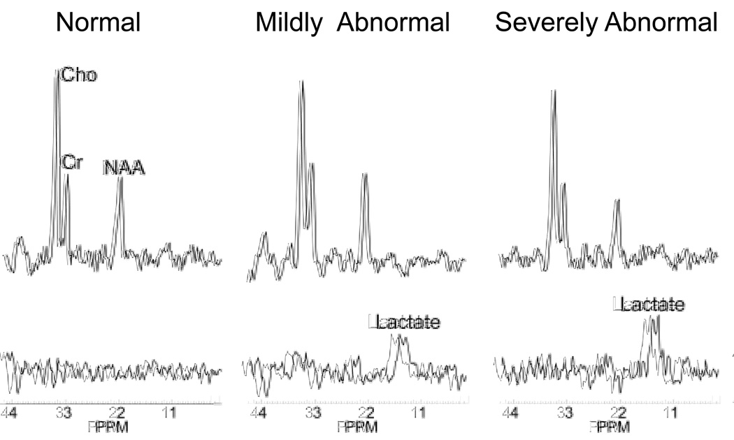
Magnetic resonance imaging has been widely used noninvasively for pediatric neuroimaging for more than a decade. More recently, with advances in computing, functional techniques for imaging water diffusion, cellular metabolite levels, and blood flow are becoming available. Magnetic resonance spectroscopy imaging (MRSI) offers a snapshot of the metabolic status in the tissue of interest. It is complementary to the more traditionally used anatomic imaging for diagnoses of various abnormalities. This review describes the physical basis of proton MRSI, summarizes currently available techniques and their applications, highlights challenges of performing MRSI in the pediatric population, and previews the newest techniques currently on the horizon.
(c) 2010 Elsevier Inc. All rights reserved.
Figures
Similar articles
-
Ultra-high resolution brain metabolite mapping at 7 T by short-TR Hadamard-encoded FID-MRSI.Neuroimage. 2018 Mar;168:199-210. doi: 10.1016/j.neuroimage.2016.10.043. Epub 2016 Nov 4. Neuroimage. 2018. PMID: 27825954
-
Magnetic resonance spectroscopy imaging (MRSI) and brain functional magnetic resonance imaging (fMRI) for radiotherapy treatment planning of glioma.Technol Cancer Res Treat. 2008 Oct;7(5):349-62. Technol Cancer Res Treat. 2008. PMID: 18783284 Review.
-
MR spectroscopic imaging: principles and recent advances.J Magn Reson Imaging. 2013 Jun;37(6):1301-25. doi: 10.1002/jmri.23945. Epub 2012 Nov 27. J Magn Reson Imaging. 2013. PMID: 23188775 Free PMC article. Review.
-
A Flow-based Truncated Denoising Diffusion Model for super-resolution Magnetic Resonance Spectroscopic Imaging.Med Image Anal. 2025 Jan;99:103358. doi: 10.1016/j.media.2024.103358. Epub 2024 Sep 27. Med Image Anal. 2025. PMID: 39353335
-
Proton MRS and MRSI of the brain without water suppression.Prog Nucl Magn Reson Spectrosc. 2015 Apr;86-87:65-79. doi: 10.1016/j.pnmrs.2014.12.001. Epub 2014 Dec 24. Prog Nucl Magn Reson Spectrosc. 2015. PMID: 25919199 Review.
Cited by
-
Brain proton magnetic resonance spectroscopy and neurodevelopment after preterm birth: a systematic review.Pediatr Res. 2022 May;91(6):1322-1333. doi: 10.1038/s41390-021-01539-x. Epub 2021 May 5. Pediatr Res. 2022. PMID: 33953356
-
Update on neuroimaging phenotypes of mid-hindbrain malformations.Neuroradiology. 2015 Feb;57(2):113-38. doi: 10.1007/s00234-014-1431-2. Epub 2014 Oct 23. Neuroradiology. 2015. PMID: 25339235
-
Cerebral magnetic resonance spectroscopy - insights into preterm brain injury.J Perinatol. 2025 Feb;45(2):194-201. doi: 10.1038/s41372-024-02172-2. Epub 2024 Nov 28. J Perinatol. 2025. PMID: 39609610 Free PMC article.
-
Impact of hypothermia on predictors of poor outcome: how do we decide to redirect care?Semin Fetal Neonatal Med. 2015 Apr;20(2):122-7. doi: 10.1016/j.siny.2014.12.011. Epub 2015 Jan 7. Semin Fetal Neonatal Med. 2015. PMID: 25577654 Free PMC article. Review.
-
Clinical 1H MRS in childhood neurometabolic diseases-part 1: technique and age-related normal spectra.Neuroradiology. 2022 Jun;64(6):1101-1110. doi: 10.1007/s00234-022-02917-w. Epub 2022 Feb 18. Neuroradiology. 2022. PMID: 35178593 Review.
References
-
- Smith FW. The Value of Nmr Imaging in Pediatric Practice - a Preliminary-Report. Pediatric Radiology. 1983;13(3):141–147. - PubMed
-
- Johnson MA, et al. Clinical Nmr Imaging of the Brain in Children - Normal and Neurologic Disease. American Journal of Neuroradiology. 1983;4(5):1013–1026. - PubMed
-
- Partridge SC, et al. Diffusion tensor imaging: serial quantitation of white matter tract maturity in premature newborns. Neuroimage. 2004;22(3):1302–1314. - PubMed
-
- Ketonen LM, Valanne L. Neuroimaging of Pediatric Diseases. Seminars in Neurology. 2008;28(4):558–569. - PubMed
Publication types
MeSH terms
Grants and funding
LinkOut - more resources
Full Text Sources
