Review
. 2010 Feb;34(1):46-56.
doi: 10.1053/j.semperi.2009.10.005.

Noninvasive cerebral perfusion imaging in high-risk neonates

Affiliations
Review

Noninvasive cerebral perfusion imaging in high-risk neonates

Donna A Goff et al. Semin Perinatol. 2010 Feb.

Abstract

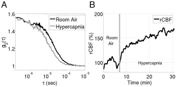
Advances in medical and surgical care of the high-risk neonate have led to increased survival. A significant number of these neonates suffer from neurodevelopmental delays and failure in school. The focus of clinical research has shifted to understanding events contributing to neurological morbidity in these patients. Assessing changes in cerebral oxygenation and regulation of cerebral blood flow (CBF) is important in evaluating the status of the central nervous system. Traditional CBF imaging methods fail for both ethical and logistical reasons. Optical near infrared spectroscopy (NIRS) is increasingly being used for bedside monitoring of cerebral oxygenation and blood volume in both very low birth weight infants and neonates with congenital heart disease. Although trends in CBF may be inferred from changes in cerebral oxygenation and/or blood volume, NIRS does not allow a direct measure of CBF in these populations. Two relatively new modalities, arterial spin-labeled perfusion magnetic resonance imaging and optical diffuse correlation spectroscopy, provide direct, noninvasive measures of cerebral perfusion suitable for the high-risk neonates. Herein we discuss the instrumentation, applications, and limitations of these noninvasive imaging techniques for measuring and/or monitoring CBF.


Figures

Figure 1
Figure 1
Labeling planes for continuous ASL (CASL) vs pulsed ASL (PASL). In CASL, continuous radio frequency (RF) is applied to the base of the brain for 1–2 seconds, magnetically labeling blood flow traveling through the plane. In PASL, RF pulses with high peak amplitude but short duration (10–20 ms) are applied to a thick area of blood inferior to the imaging plane. (Color version of figure is available online.)
Figure 2
Figure 2
Direct comparison of mean whole brain CBF as measured by PCASL (y-axis) and PASL (x-axis).
Figure 3
Figure 3
A direct comparison of PASL (upper) and pseudo CASL (lower). Images were obtained sequentially in a single patient both at rest (baseline) and hypercarbia. The 2 right arrows demonstrate increased negative pixels on PASL imaging in the left frontal cortex. The arrowheads on the left demonstrate improved anatomic resolution in the central sulcus. (Color version of figure is available online.)
Figure 4
Figure 4
Direct comparison of PCASL and PASL perfusion magnetic resonance imaging (MRI) in a cohort of infants and children with congenital heart disease. With increased values of cerebral blood flow (CBF), the percentage of negative voxels is reduced. PCASL offers an improvement in SNR at low blood flow values.
Figure 5
Figure 5
Comparison of PASL perfusion MRI at 1.5 T (closed triangles) and 3 T (open squares) along with the best fit lines to both sets of data. Patients studied on the 3 T were all full-term infants with congenital heart defects, imaged before surgery in the first week of life.
Figure 6
Figure 6
A sample absorption spectrum of oxyhemoglobin, deoxyhemoglobin, and water in tissue. A spectral “window” exists in the near-infrared range (highlighted in the gray box and enlarged above), so that scattering, rather than absorption, dominates photon propagation. (Color version of figure is available online.)
Figure 7
Figure 7
Depiction of photon propagation through brain tissue. Light that reaches the detector is most likely affected by tissue in the darkest red region, while regions in orange or yellow have less effect on the photons’ journey. The depth of photon penetration is highly dependent on source-detector separation. Larger separations lead to deeper penetration. (Color version of figure is available online.)
Figure 8
Figure 8
Graphical depiction of the 3 types of near infrared spectroscopy measurements: (A) Continuous wave, (B) frequency domain, and (C) time resolved. The arrows on the right side of the figure indicate the increasing data and instrumentation complexity which accompany these techniques. (Color version of figure is available online.)
Figure 9
Figure 9
(A) sample autocorrelation curve measured with diffuse correlation spectroscopy on a neonate with hypoplastic left heart syndrome. The “baseline” curve, taken on the patient’s forehead during room air inhalation, decays slower than the hypercapnia curve taken while the patient inhaled a CO2 gas mixture. The increase in the decay rate during hypercapnia indicates an increase in CBF compared with the resting baseline period. (B) Trace of relative change in cerebral blood flow (rCBF) during this hypercapnia experiment extracted from autocorrelation curves measured every 3 seconds.

Similar articles

Cited by

References

    1. Mathur A, Inder T. Magnetic resonance imaging—insights into brain injury and outcomes in premature infants. J Commun Disord. 2009;42:248–255. - PMC - PubMed
    1. Back SA, Han BH, Luo NL, et al. Selective vulnerability of late oligodendrocyte progenitors to hypoxia-ischemia. J Neurosci. 2002;22:455–463. - PMC - PubMed
    1. Drobyshevsky A, Song SK, Gamkrelidze G, et al. Developmental changes in diffusion anisotropy coincide with immature oligodendrocyte progression and maturation of compound action potential. J Neurosci. 2005;25:5988–5997. - PMC - PubMed
    1. Back SA, Riddle A, McClure MM. Maturation-dependent vulnerability of perinatal white matter in premature birth. Stroke. 2007;38:724–730. - PubMed
    1. Rezaie P, Dean A. Periventricular leukomalacia, inflammation and white matter lesions within the developing nervous system. Neuropathology. 2002;22:106–132. - PubMed

Publication types

MeSH terms