

Fluorescent probes for the analysis of DNA strand scission in base excision repair
- PMID: 20110254
- PMCID: PMC2853145
- DOI: 10.1093/nar/gkq022
Fluorescent probes for the analysis of DNA strand scission in base excision repair
Abstract
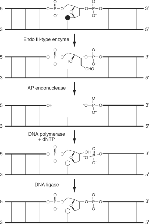
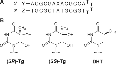
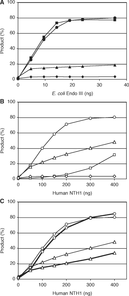
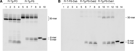
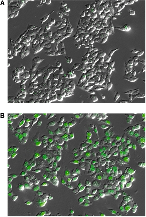
We have developed fluorescent probes for the detection of strand scission in the excision repair of oxidatively damaged bases. They were hairpin-shaped oligonucleotides, each containing an isomer of thymine glycol or 5,6-dihydrothymine as a damaged base in the center, with a fluorophore and a quencher at the 5'- and 3'-ends, respectively. Fluorescence was detected when the phosphodiester linkage at the damage site was cleaved by the enzyme, because the short fragment bearing the fluorophore could not remain in a duplex form hybridized to the rest of the molecule at the incubation temperature. The substrate specificities of Escherichia coli endonuclease III and its human homolog, NTH1, determined by using these probes agreed with those determined previously by gel electrophoresis using (32)P-labeled substrates. Kinetic parameters have also been determined by this method. Since different fluorophores were attached to the oligonucleotides containing each lesion, reactions with two types of substrates were analyzed separately in a single tube, by changing the excitation and detection wavelengths. These probes were degraded during an incubation with a cell extract. Therefore, phosphorothioate linkages were incorporated to protect the probes from nonspecific nucleases, and the base excision repair activity was successfully detected in HeLa cells.
Figures
Similar articles
-
Fluorescence detection of the endonuclease III reaction using modified oligonucleotides.Nucleic Acids Symp Ser (Oxf). 2009;(53):213-4. doi: 10.1093/nass/nrp107. Nucleic Acids Symp Ser (Oxf). 2009. PMID: 19749336
-
Excision of 5,6-dihydroxy-5,6-dihydrothymine, 5,6-dihydrothymine, and 5-hydroxycytosine from defined sequence oligonucleotides by Escherichia coli endonuclease III and Fpg proteins: kinetic and mechanistic aspects.Biochemistry. 1999 Mar 16;38(11):3335-44. doi: 10.1021/bi981982b. Biochemistry. 1999. PMID: 10079077
-
Comparison of substrate specificities of Escherichia coli endonuclease III and its mouse homologue (mNTH1) using defined oligonucleotide substrates.Biochemistry. 2000 Sep 19;39(37):11389-98. doi: 10.1021/bi000422l. Biochemistry. 2000. PMID: 10985784
-
Single-Labeled Oligonucleotides Showing Fluorescence Changes Upon Hybridization with Target Nucleic Acids.Molecules. 2018 Jan 8;23(1):124. doi: 10.3390/molecules23010124. Molecules. 2018. PMID: 29316733 Free PMC article. Review.
-
DNA substrates containing defined oxidative base lesions and their application to study substrate specificities of base excision repair enzymes.Prog Nucleic Acid Res Mol Biol. 2001;68:207-21. doi: 10.1016/s0079-6603(01)68101-7. Prog Nucleic Acid Res Mol Biol. 2001. PMID: 11554298 Review.
Cited by
-
Fluorescence detection of DNA mismatch repair in human cells.Sci Rep. 2018 Aug 15;8(1):12181. doi: 10.1038/s41598-018-30733-x. Sci Rep. 2018. PMID: 30111891 Free PMC article.
-
Fluorescent Probes of DNA Repair.ACS Chem Biol. 2018 Jul 20;13(7):1721-1733. doi: 10.1021/acschembio.7b00919. Epub 2017 Nov 30. ACS Chem Biol. 2018. PMID: 29156135 Free PMC article. Review.
-
Chimeric d/l-DNA Probes of Base Excision Repair Enable Real-Time Monitoring of Thymine DNA Glycosylase Activity in Live Cells.J Am Chem Soc. 2023 Aug 9;145(31):17066-17074. doi: 10.1021/jacs.3c03010. Epub 2023 Jul 26. J Am Chem Soc. 2023. PMID: 37493592 Free PMC article.
-
NEIL1 binding to DNA containing 2'-fluorothymidine glycol stereoisomers and the effect of editing.Chembiochem. 2012 Jun 18;13(9):1338-48. doi: 10.1002/cbic.201200139. Epub 2012 May 25. Chembiochem. 2012. PMID: 22639086 Free PMC article.
-
Fluorescence detection of cellular nucleotide excision repair of damaged DNA.Sci Rep. 2014 Jul 4;4:5578. doi: 10.1038/srep05578. Sci Rep. 2014. PMID: 24993089 Free PMC article.
References
-
- Wilson DM, Bohr VA. The mechanics of base excision repair, and its relationship to aging and disease. DNA Repair. 2007;6:544–559. - PubMed
-
- Rechkunova NI, Maltseva EA, Lavrik OI. Nucleotide excision repair in higher eukaryotes: mechanism of primary damage recognition. Mol. Biol. 2008;42:20–26. - PubMed
-
- Leibeling D, Laspe P, Emmert S. Nucleotide excision repair and cancer. J. Mol. Hist. 2006;37:225–238. - PubMed
Publication types
MeSH terms
Substances
LinkOut - more resources
Full Text Sources
Molecular Biology Databases
