. 2010 Jun;38(10):3328-39.
doi: 10.1093/nar/gkq018. Epub 2010 Jan 27.

Evolutionary optimization of a modular ligase ribozyme: a small catalytic unit and a hairpin motif masking an element that could form an inactive structure

Affiliations

Evolutionary optimization of a modular ligase ribozyme: a small catalytic unit and a hairpin motif masking an element that could form an inactive structure

Yuki Fujita et al. Nucleic Acids Res. 2010 Jun.

Abstract

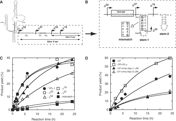
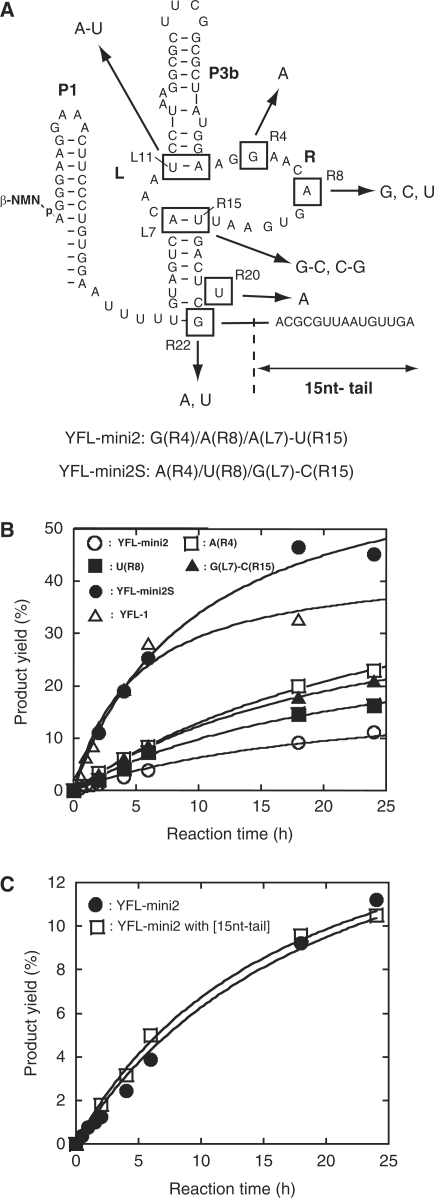
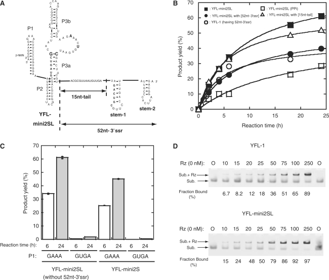
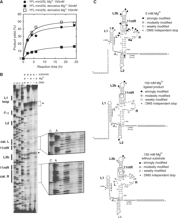
The YFL ribozyme is an artificial ligase ribozyme isolated by a 'design and selection' strategy, in which a modular catalytic unit was generated on a rationally designed modular scaffold RNA. This ligase ribozyme has a versatile catalytic unit that accepts not only beta-nicotinamide mononucleotide (beta-NMN) but also inorganic pyrophosphate as leaving groups for template-dependent RNA ligation. Although this property is interesting from an evolutionary viewpoint regarding primitive RNA ligation/polymerization systems in the RNA world, structural analysis of the YFL ribozyme has not been continued due to apparent structural nonuniformity of its folded state. To elucidate the active structure of the YFL ribozyme, we performed in vitro evolution experiments to improve its folding ability. Biochemical and phylogenetic analyses of evolved variants indicated that the catalytic unit of the YFL ribozyme is compact and the 3' single-stranded region of the parent YFL-1 ribozyme contributes to mask an element that could form an inactive structure.


Figures

Figure 1.
Figure 1.
Construction and miniaturization of the class YFL ribozyme. (A) Generation and miniaturization of the class YFL ribozyme. The YFL-1 ribozyme (middle) was constructed by installing catalytic units into the Type B scaffold (left). YFL-mini1 (right) is a truncated variant lacking regions dispensable for minimal catalytic ability. In the structure of YFL-1 (middle), the arrow with a black arrowhead indicates the site downstream of which was removed in the Δ25 mutant. In the structure of YFL-mini1 (right), the nucleotide numbering for the catalytic unit and its surrounding elements is shown in red. (B) Time courses of the ligation reactions by the YFL-1 (filled circles) and its Δ25 mutant lacking the 25-nt-3′ssr (open squares). Reactions were performed in the presence of 30 mM Tris–HCl (pH 7.5), 50 mM MgCl2 and 200 mM KCl at 37°C. (C) Product yields of YFL-1 with the 25-nt-3′ssr, its variants lacking part of the 25-nt-3′ssr, and the Δ25 mutant lacking the whole 25-nt-3′ssr.
Figure 2.
Figure 2.
In vitro evolution of the YFL-mini1 ribozyme. (A) Starting pool designed based on YFL-mini1. Five nucleotides in the L region were replaced with random nucleotides (N). The R region was partially mutagenized with 9% or 21% degeneracy (lowercase letters). (B) Scheme for in vitro evolution to identify variants, activity of which is independent of the 3′-end sequence. (1) The RNA pool was incubated with a biotinylated substrate and active variants were selectively retained on streptavidin magnetic beads. (2) Active variants were preferentially amplified by reverse transcription. (3) After degradation of RNA, ( and 5) cDNAs complementary to active variants were amplified by PCR. (6) The 3′ Tag region of resulting DNAs, corresponding to the 3′ssr for RT-PCR in the RNA sequence, was digested with BanI. (7) An alternate Tag sequence was attached by T4 ligase. (8) The resulting DNAs were transcribed and used for the next round.
Figure 3.
Figure 3.
Sequence comparison of YFL-mini1 and its variants isolated by in vitro evolution. (A) Sequences of 21 clones from the pool after the 5th round. Dashes indicate the bases identical to those of the parent YFL-mini1. (B) Base substitutions found in the isolated clones were mapped on the originally proposed structure (structure-1) of YFL-mini1. (C) The revised structure model derived from the phylogenetic comparison of the isolated clones (structure-2).
Figure 4.
Figure 4.
Examination of catalytic abilities of structures-1 and -2. (A, B) Secondary structures of YFL-mini1 and its mutants. mut-1 (A) was designed to form the originally proposed structure (structure-1). mut-2c (B) was designed to form the revised structure (structure-2). mut-2a and mut-2b (B) were designed to disrupt the stem region of structure-2. (C) Reactions of YFL-1, YFL-mini1 and the four mutants of YFL-mini1. Reactions were performed with 30 mM Tris–HCl (pH 7.5), 100 mM Mg2+, 200 mM K+ at 37°C. ‘YFL-1-sub’. indicates the ligated product of the YFL-1 ribozyme and the substrate oligonucleotide. ‘Product’ indicates the ligated product of the substrate and the YFL-mini1 or its mutant. The asterisk indicates the product presumably formed by specific hydrolytic cleavage of the ligated product of YFL-1, which should be promoted by the RNA tertiary structure (22).
Figure 5.
Figure 5.
Possible stem structure in the 52-nt-3′ssr. Sequence (A) and possible secondary structure (B) of 52-nt-3′ssr. The secondary structure was predicted using the mfold program. Arrows with black arrowheads indicate the truncation positions. (C) Activities of the parent YFL-1 and its mutants to examine the effects of 52-nt-3′ssr. (D) Activities of the parent YFL-1 and its mutants to examine the effects of the 15-nt-tail. Δ37–comp. oligo indicates the reaction of the Δ37 mutant in the presence of the indicated concentrations of a DNA oligonucleotide complementary to the 15-nt-tail region.
Figure 6.
Figure 6.
Effects of mutations in the selected clones. (A) Mutations isolated by in vitro evolution. They were categorized into six positions in a shortened form of the YFL-1 (YFL-mini2). The YFL-mini2S mutant is the most active mutant of YFL-mini2, which possesses two nucleotide substitutions [G(R4)A and A(R8)U] and a base pair substitution [A(L7)G-U(R15)C]. (B) Activities of YFL-mini2 and its variants lacking the 15-nt tail. (C) Activities of the YFL-mini2 and its mutant with the 15-nt tail.
Figure 7.
Figure 7.
Effects of 3′ssr on the YFL-mini2SL ribozyme. (A) Schematic representation of the secondary structure of YFL-mini2SL with or without the 52-nt-3′ssr. (B) Time courses of ligation reactions catalyzed by YFL-1, YFL-mini2SL and YFL-mini2SL variants. YFL-mini2SL (PPi) indicates a variant of YFL-mini2SL that uses inorganic pyrophosphate (PPi) as a leaving group. (C) Activities of the YFL-mini2SL and YFL-mini2S variants with mutations in the P1 loop. (D) Gel mobility shift assays of YFL-1 or YFL-mini2SL with the substrates in the presence of 50 mM Mg2+ ions at pH 7.5. The FAM-labeled substrate unit the 3′-end of which has deoxyribose (25 nM) was titrated with various concentrations of the ribozyme (0–250 nM). Rz and Sub. indicate the ribozyme and substrate, respectively. Upper and lower bands correspond to the substrate–ribozyme complex (Sub. + Rz) and free substrate (Sub.), respectively. Experiments were performed according to the published protocol (23).
Figure 8.
Figure 8.
DMS modification of a derivative of YFL-mini2SL. (A) Time courses of the ligation reactions of YFS-mini2SL and its variant for DMS analysis. (B) DMS modification of a derivative of YFL-mini2SL. Asterisks indicate the stops of reverse transcription that occur independently from DMS modification. (C) Positions and extents of DMS modification in the absence (top) or presence (middle and bottom) of 150 mM MgCl2. In the absence of MgCl2, DMS modification was performed with the ribozyme in the presence of the substrate. In the presence of 150 mM MgCl2, DMS modification was performed with either the ligated product produced in situ (middle) or with the ribozyme in the absence of the substrate (bottom).

Similar articles

Cited by

References

    1. Campbell ID, Baron M. The structure and function of protein modules. Philos. Trans. R. Soc. Lond. B Biol. Sci. 1991;332:165–170. - PubMed
    1. Campbell ID, Downing AK. Building protein structure and function from modular units. Trends Biotechnol. 1994;12:168–172. - PubMed
    1. Brion P, Westhof E. Hierarchy and dynamics of RNA folding. Annu. Rev. Biophys. Biomol. Struct. 1997;26:113–137. - PubMed
    1. Jaeger L. The new world of ribozymes. Curr. Opin. Struct. Biol. 1997;7:324–335. - PubMed
    1. Westhof E, Masquida B, Jaeger L. RNA tectonics: towards RNA design. Fold. Des. 1996;1:R78–R88. - PubMed

Publication types