. 2010 Mar;137(5):755-65.
doi: 10.1242/dev.045757. Epub 2010 Jan 28.

SIX1 acts synergistically with TBX18 in mediating ureteral smooth muscle formation

Affiliations

SIX1 acts synergistically with TBX18 in mediating ureteral smooth muscle formation

Xuguang Nie et al. Development. 2010 Mar.

Abstract

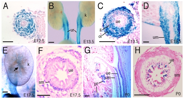
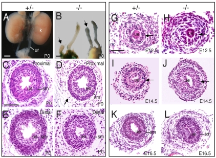
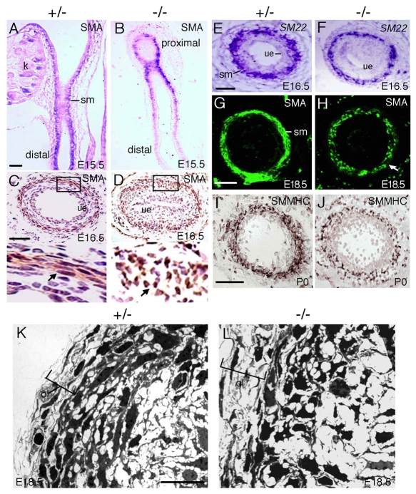
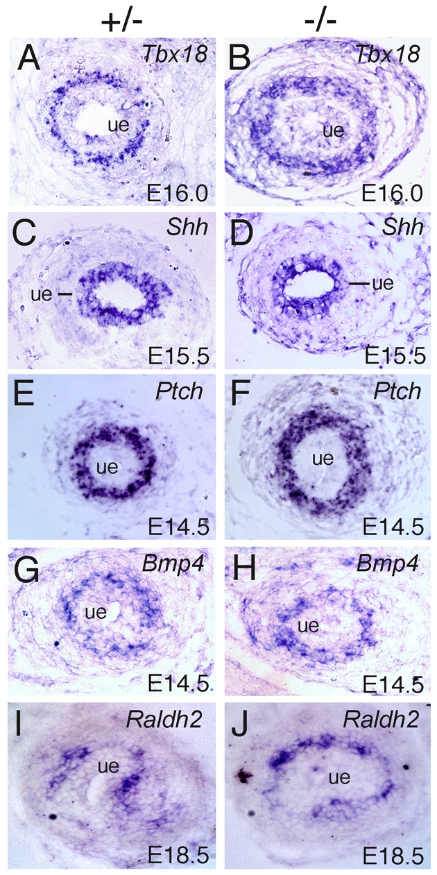
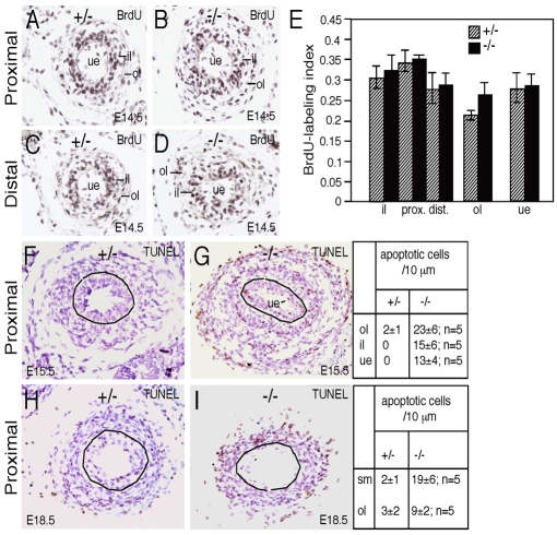
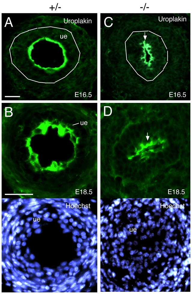
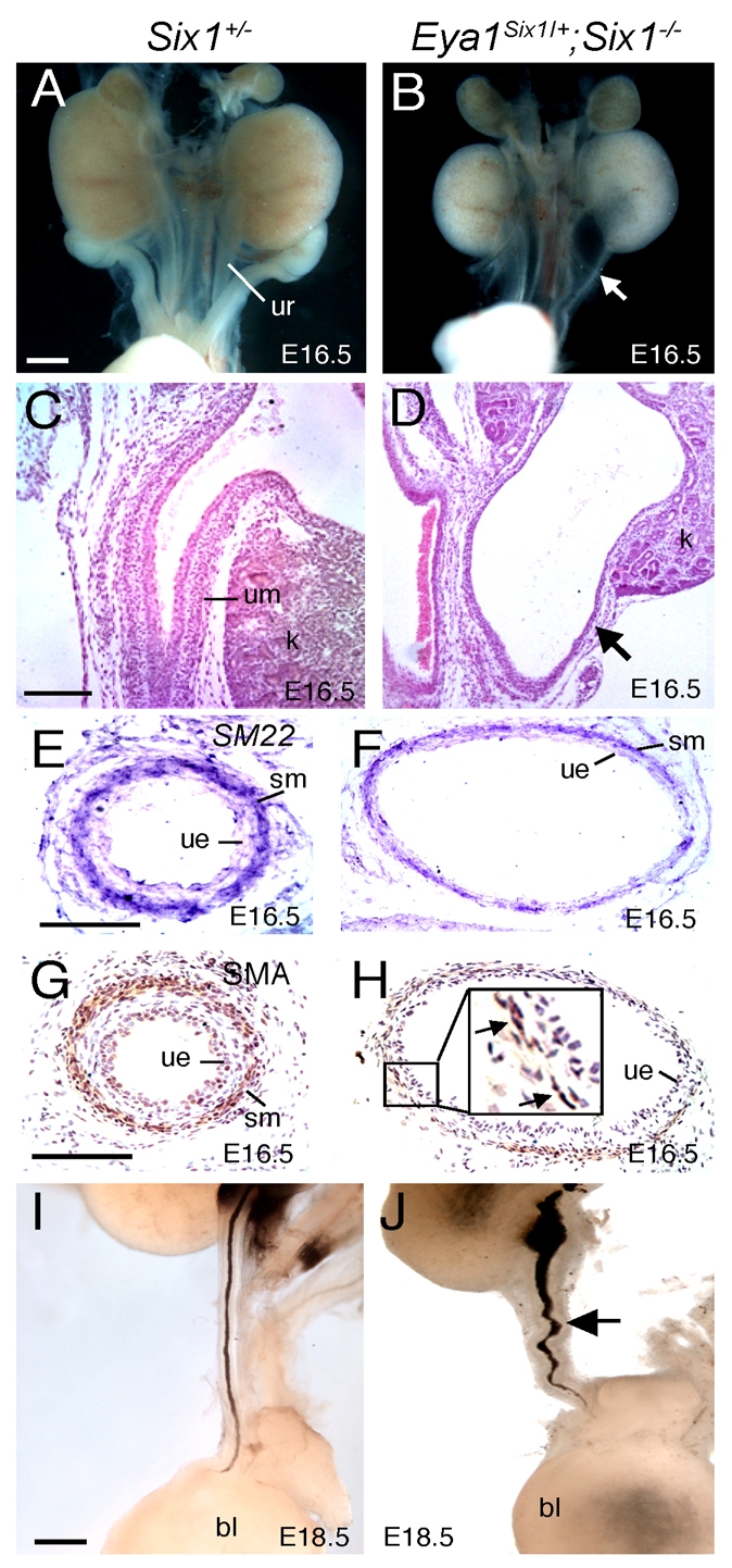
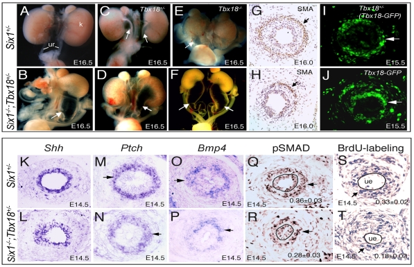
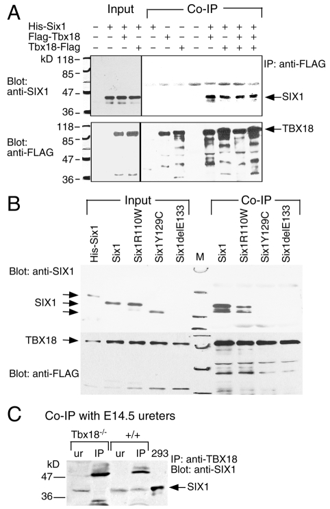
Dysfunction of the ureter often leads to urine flow impairment from the kidney to the bladder, causing dilation of the ureter and/or renal pelvis. Six1 is a crucial regulator of renal development: mutations in human SIX1 cause branchio-oto-renal (BOR) syndrome and Six1(-/-) mice exhibit renal agenesis, although the ureter is present. It remains unclear whether Six1 plays a role in regulating ureter morphogenesis. We demonstrate here that Six1 is differentially expressed during ureter morphogenesis. It was expressed in undifferentiated smooth muscle (SM) progenitors, but was downregulated in differentiating SM cells (SMCs) and had disappeared by E18.5. In Six1(-/-) mice, the ureteral mesenchymal precursors failed to condense and differentiate into normal SMCs and showed increased cell death, indicating that Six1 is required for the maintenance and normal differentiation of SM progenitors. A delay in SMC differentiation was observed in Six1(-/-) ureters. A lack of Six1 in the ureter led to hydroureter and hydronephrosis without anatomical obstruction when kidney formation was rescued in Six1(-/-) embryos by specifically expressing Six1 in the metanephric mesenchyme, but not the ureter, under control of the Eya1 promoter. We show that Six1 and Tbx18 genetically interact to synergistically regulate SMC development and ureter function and that their gene products form a complex in cultured cells and in the developing ureter. Two missense mutations in SIX1 from BOR patients reduced or abolished SIX1-TBX18 complex formation. These findings uncover an essential role for Six1 in establishing a functionally normal ureter and provide new insights into the molecular basis of urinary tract malformations in BOR patients.


Figures

Fig. 1.
Fig. 1.
Expression of Six1 in the developing mouse ureter. Sections are X-gal stained for lacZ expression (blue). (A) A transverse section of E12.5 Six1+/lacZ ureter. (B) Whole-mount Six1+/lacZ kidney (k) and ureter (ur). (C) A transverse section of the ureter shown in B. (D) A longitudinal section of E15.5 Six1+/lacZ ureter. (E) Whole-mount E17.5 Six1+/lacZ kidney and ureter. (F) A transverse section of the ureter shown in E. (G) A longitudinal section of P0 Six1+/lacZ kidney and ureter. (H) A transverse section of the P0 Six1+/lacZ ureter. il, inner mesenchymal layer; ol, outer mesenchymal layer; pt, proximal tubules; sm, smooth muscle layer; ue, urothelium; um, ureteral mesenchyme. Scale bars: 50 μm.
Fig. 2.
Fig. 2.
Ureter anomalies in Six1–/– embryos. (A,B) Urinary tract systems in Six1+/– control (A) and Six1–/– (B) mouse embryos. Arrows point to truncation of the ureters (ur). B shows X-gal-stained (left) and unstained (right) urinary tract systems. (C-F) Transverse sections showing the proximal and distal regions of the Six1+/– (C,E) and Six1–/– (D,F) ureter. (G-L) Transverse sections of ureters in Six1+/– (G,I,K) and littermate Six1–/– (H,J,L) embryos from E12.5-16.5. Arrows point to condensing mesenchyme that forms the smooth muscle (sm) layer. ue, ureteric epithelium. Scale bars: 50 μm.
Fig. 3.
Fig. 3.
Ureteric SMC differentiation in Six1+/– control and Six1–/– mouse ureters. (A,B) Longitudinal sections of the ureter showing SMA in the differentiating SMCs (detected using BCIP/NBT). (C,D) Transverse sections showing SMA staining (detected using DAB). The lower panels of C and D show higher magnification images of the boxed areas. Arrows point to spindle-shaped SMCs in control ureters and disoriented SMCs in Six1–/– ureters. (E,F) Transverse sections of proximal ureters showing SM22α expression in differentiating SMCs. (G,H) Transverse sections showing SMA staining. Arrow points to disoriented SMCs. (I,J) Transverse sections showing SMMHC staining. (K,L) Transmission electron micrographs of E18.5 ureter transverse sections. ol, outer layer of the ureteral mesenchyme. Scale bars: 50 μm in A-J; 10 μm in K,L.
Fig. 4.
Fig. 4.
Molecular marker analysis of Six1–/– ureteral mesenchyme. In situ hybridization on transverse sections of Six1+/– control and Six1–/– mouse ureters with (A,B) Tbx18, (C,D) Shh, (E,F) Ptch1, (G,H) Bmp4 and (I,J) Raldh2. ue, ureteric epithelium.
Fig. 5.
Fig. 5.
Cell proliferation and apoptosis in Six1–/– ureters. (A-D) Cell proliferation analysis by BrdU staining on E14.5 Six1+/– control and Six1–/– mouse ureter transverse sections. (E) Statistical analysis of BrdU-positive cells. BrdU-positive cells and the total number of cells in each layer on each transverse section were counted and data from six sections from each ureter (from four ureters) were normalized as the BrdU labeling index. In the mesenchyme: P=0.1361, control inner layer (il) (0.31±0.03) versus mutant inner layer (0.323±0.04); P=0.0325, control proximal inner layer (prox.) (0.34±0.03) versus mutant proximal inner layer (0.35±0.01); P=0.0782, control distal inner layer (dist.) (0.28±0.04) versus mutant distal inner layer (0.29±0.03); P=0.0964, control outer layer (ol) (0.23±0.01) versus mutant outer layer (0.27±0.02). In E14.5 ureteric epithelium (ue): P=0.1927, control (0.28±0.04) versus mutant (0.29±0.03). P-values were calculated using StatView t-test; error bars indicate s.d. (F-I) TUNEL analysis of E15.5 and E18.5 Six1+/– control and Six1–/– ureters. TUNEL-positive cells (brown nuclei) were counted from five serial sections from each ureter (from at least six ureters). Shown are the average number of TUNEL-positive cells per (10 μm) section for each genotype. sm, smooth muscle layer.
Fig. 6.
Fig. 6.
Uroplakin expression in control and mutant ureters. Immunostaining for uroplakin IIIa (green) in urothelium (ue) of (A,B) Six1+/– and (C,D) Six1–/–mouse embryos. Lower panels of B and D are counter-stained with Hoechst. Scale bars: 50 μm.
Fig. 7.
Fig. 7.
Lack of Six1 in the ureter leads to hydroureter and hydronephrosis. (A,B) Whole E16.5 urinary tracts from Six1+/– and Eya1Six1/+;Six1–/– mouse embryos. Arrow points to hydroureter in Eya1Six1/+;Six1–/–. (C,D) Hematoxylin and Eosin-stained longitudinal sections of the kidney (k) and ureters shown in A and B. Arrow indicates the thin wall of ureteral mesenchyme (um) in the mutant. (E-H) In situ hybridization for SM22α (E,F) and immunostaining for SMA (G,H) on ureter transverse sections. Arrows in the higher magnification image in H point to SMCs. (I,J) E18.5 urinary tracts in which India ink was injected into the renal pelvis. Arrow points to dilated ureter in the Eya1Six1/+;Six1–/– ureter. bl, bladder; ue, urothelium. Scale bars: 50 μm.
Fig. 8.
Fig. 8.
Six1 and Tbx18 genetically interact during ureter development. (A-F) Urogenital systems in (A) Six1+/–, (B,D,F) Six1–/–;Tbx18+/–, (C) Six1+/–;Tbx18–/– and (E) Six1–/–;Tbx18–/– mouse embryos. Arrows point to dilated ureters. Note the severe dilation in Six1–/–;Tbx18+/– embryos. (G,H) Immunostaining for SMA on Six1+/– (G) and Six1–/–;Tbx18+/– (H) transverse sections. (I,J) Transverse sections of ureters of Six1+/–;Tbx18+/– (Tbx18+/GFP) and Six1–/–;Tbx18+/– embryos showing Tbx18-GFP expression in the ureteral mesenchyme. Arrows indicate condensing progenitors. Note that more Tbx18-GFP-positive cells were distributed in the outer layer in the Six1–/–;Tbx18+/– mutant than in the Six1+/–;Tbx18+/– control ureters. (K-P) In situ hybridization for Shh, Ptch1 and Bmp4 on transverse sections. (Q,R) Immunohistochemistry for pSMAD on transverse sections from Six1+/– and Six1–/–;Tbx18+/– embryos. Arrows point to pSMAD-positive nuclei in the condensing mesenchyme. pSMAD-positive cells from five sections from each ureter (three ureters) were quantified as the ratio of pSMAD-positive cells to the total number of condensing mesenchymal cells in the control and mutant ureters. P-values (P=0.1039) were calculated using StatView t-test. (S,T) Transverse sections showing BrdU-positive cells (arrows). Proliferative rate was quantified as the ratio of BrdU-labeled cells to the total number of cells in the condensing mesenchyme from six sections from each ureter (from at least four ureters) on a radial plane. P-values (P=0.003) were calculated using StatView t-test. ue, urothelium.
Fig. 9.
Fig. 9.
SIX1 and TBX18 physically interact in vitro and in vivo. (A) Western blot and co-IP. Cell extracts (5 μg) from HEK 293 cells transfected with different combinations of His-Six1/pcDNA3, Flag-Tbx18 or Tbx18-Flag were separated in SDS-PAGE gels and analyzed by western blotting with anti-SIX1 or anti-FLAG antibody to detect HIS-SIX1, FLAG-TBX18 or TBX18-FLAG. For co-IP experiments, ∼0.4 mg of the same extracts was used. (B) Western blot and co-IP. Cell extracts (0.4 mg) from HEK 293 cells co-transfected with His-Six1/pcDNA3, Six1/pcDNA, its missense mutants (R110W, Y129C) or deletion mutant (delE133) and Flag-Tbx18 were separated in SDS-PAGE gels and analyzed by western blotting with anti-SIX1 or anti-FLAG antibody to detect SIX1 or FLAG-TBX18.The antibody failed to detect the mutant delE133 protein. (C) Western blot and co-IP. Cell extracts (∼3 mg) from E14.5 ureters were incubated with anti-TBX18 and precipitated by protein G-agarose beads. The precipitates were dissolved in SDS sample buffer followed by western blot analysis with anti-SIX1. ur, ureter; 293, cell lysates from HEK 293 cells transfected with Flag-Tbx18 and Six1.

Similar articles

Cited by

References

    1. Abdelhak S., Kalatzis V., Heilig R., Compain S., Samson D., Vincent C., Weil D., Cruaud C., Sahly I., Leibovici M., et al. (1997). A human homologue of the Drosophila eyes absent gene underlies branchio-oto-renal (BOR) syndrome and identifies a novel gene family. Nat. Genet. 15, 157-164 - PubMed
    1. Airik R., Bussen M., Singh M. K., Petry M., Kispert A. (2006). Tbx18 regulates the development of the ureteral mesenchyme. J. Clin. Invest. 116, 663-674 - PMC - PubMed
    1. Brenner-Anantharam A., Cebrian C., Guillaume R., Hurtado R., Sun T. T., Herzlinger D. (2007). Tailbud-derived mesenchyme promotes urinary tract segmentation via BMP4 signaling. Development 134, 1967-1975 - PubMed
    1. Buller C., Xu X., Marquis V., Schwanke R., Xu P. X. (2001). Molecular effects of Eya1 domain mutations causing organ defects in BOR syndrome. Hum. Mol. Genet. 10, 2775-2781 - PubMed
    1. Bush K. T., Vaughn D. A., Li X., Rosenfeld M. G., Rose D. W., Mendoza S. A., Nigam S. K. (2006). Development and differentiation of the ureteric bud into the ureter in the absence of a kidney collecting system. Dev. Biol. 298, 571-584 - PubMed

Publication types

MeSH terms