BMP signaling induces digit regeneration in neonatal mice
- PMID: 20110320
- PMCID: PMC2827613
- DOI: 10.1242/dev.042424
BMP signaling induces digit regeneration in neonatal mice
Abstract
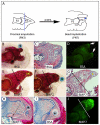
The regenerating digit tip of mice is a novel epimorphic response in mammals that is similar to fingertip regeneration in humans. Both display restricted regenerative capabilities that are amputation-level dependent. Using this endogenous regeneration model in neonatal mice, we have found that noggin treatment inhibits regeneration, thus suggesting a bone morphogenetic protein (BMP) requirement. Using non-regenerating amputation wounds, we show that BMP7 or BMP2 can induce a regenerative response. BMP-induced regeneration involves the formation of a mammalian digit blastema. Unlike the endogenous regeneration response that involves redifferentiation by direct ossification (evolved regeneration), the BMP-induced response involves endochondral ossification (redevelopment). Our evidence suggests that BMP treatment triggers a reprogramming event that re-initiates digit tip development at the amputation wound. These studies demonstrate for the first time that the postnatal mammalian digit has latent regenerative capabilities that can be induced by growth factor treatment.
Figures


Similar articles
-
SDF-1α/CXCR4 signaling mediates digit tip regeneration promoted by BMP-2.Dev Biol. 2013 Oct 1;382(1):98-109. doi: 10.1016/j.ydbio.2013.07.020. Epub 2013 Aug 2. Dev Biol. 2013. PMID: 23916851
-
Temporal requirement for bone morphogenetic proteins in regeneration of the tail and limb of Xenopus tadpoles.Mech Dev. 2006 Sep;123(9):674-88. doi: 10.1016/j.mod.2006.07.001. Epub 2006 Jul 6. Mech Dev. 2006. PMID: 16938438
-
BMP2 induces segment-specific skeletal regeneration from digit and limb amputations by establishing a new endochondral ossification center.Dev Biol. 2012 Dec 15;372(2):263-73. doi: 10.1016/j.ydbio.2012.09.021. Epub 2012 Oct 3. Dev Biol. 2012. PMID: 23041115 Free PMC article.
-
Evolution of epimorphosis in mammals.J Exp Zool B Mol Dev Evol. 2021 Mar;336(2):165-179. doi: 10.1002/jez.b.22925. Epub 2020 Jan 17. J Exp Zool B Mol Dev Evol. 2021. PMID: 31951104 Review.
-
[Application of BMP to bone repair].Clin Calcium. 2007 Feb;17(2):263-9. Clin Calcium. 2007. PMID: 17272885 Review. Japanese.
Cited by
-
Cellular and molecular mechanisms that regulate mammalian digit tip regeneration.Open Biol. 2020 Sep;10(9):200194. doi: 10.1098/rsob.200194. Epub 2020 Sep 30. Open Biol. 2020. PMID: 32993414 Free PMC article.
-
Extracellular vesicles derived from Msh homeobox 1 (Msx1)-overexpressing mesenchymal stem cells improve digit tip regeneration in an amputee mice model.Sci Rep. 2024 Oct 9;14(1):23538. doi: 10.1038/s41598-024-72647-x. Sci Rep. 2024. PMID: 39384602 Free PMC article.
-
Advances in understanding tissue regenerative capacity and mechanisms in animals.Nat Rev Genet. 2010 Oct;11(10):710-22. doi: 10.1038/nrg2879. Epub 2010 Sep 14. Nat Rev Genet. 2010. PMID: 20838411 Free PMC article. Review.
-
A pivotal role for Wnt antagonists in constraining Wnt activity to promote digit joint specification.bioRxiv [Preprint]. 2025 Jul 21:2025.07.17.665381. doi: 10.1101/2025.07.17.665381. bioRxiv. 2025. PMID: 40747425 Free PMC article. Preprint.
-
Potassium channels as potential drug targets for limb wound repair and regeneration.Precis Clin Med. 2020 Mar;3(1):22-33. doi: 10.1093/pcmedi/pbz029. Epub 2019 Dec 30. Precis Clin Med. 2020. PMID: 32257531 Free PMC article.
References
-
- Ahn K., Mishina Y., Hanks M. C., Behringer R. R., Crenshaw E. B., 3rd (2001). BMPR-IA signaling is required for the formation of the apical ectodermal ridge and dorsal-ventral patterning of the limb. Development 128, 4449-4461 - PubMed
-
- Allan C. H., Fleckman P., Fernandes R. J., Hager B., James J., Wisecarver Z., Satterstrom F. K., Gutierrez A., Norman A., Pirrone A., et al. (2006). Tissue response and Msx1 expression after human fetal digit tip amputation in vitro. Wound Repair Regen. 14, 398-404 - PubMed
-
- Brockes J. P., Kumar A. (2005). Appendage regeneration in adult vertebrates and implications for regenerative medicine. Science 310, 1919-1923 - PubMed
-
- Bryant S. V., Endo T., Gardiner D. M. (2002). Vertebrate limb regeneration and the origin of limb stem cells. Int. J. Dev. Biol. 46, 887-896 - PubMed
Publication types
MeSH terms
Substances
Grants and funding
LinkOut - more resources
Full Text Sources
Other Literature Sources

