

Notch signalling regulates the contribution of progenitor cells from the chick Hensen's node to the floor plate and notochord
- PMID: 20110321
- PMCID: PMC3928719
- DOI: 10.1242/dev.041608
Notch signalling regulates the contribution of progenitor cells from the chick Hensen's node to the floor plate and notochord
Abstract
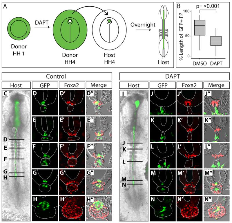
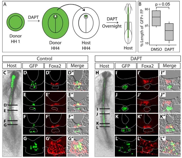
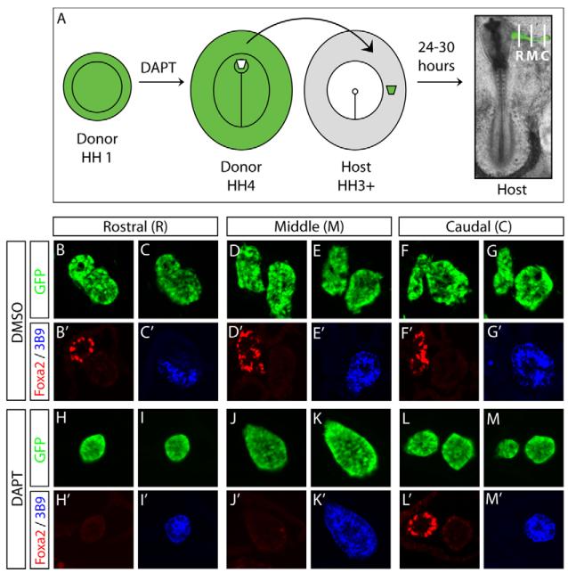
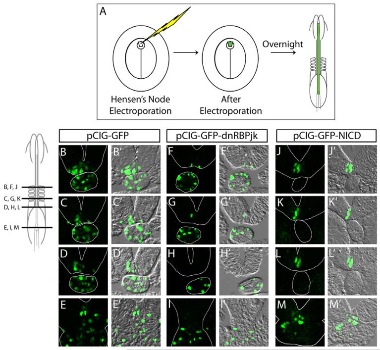
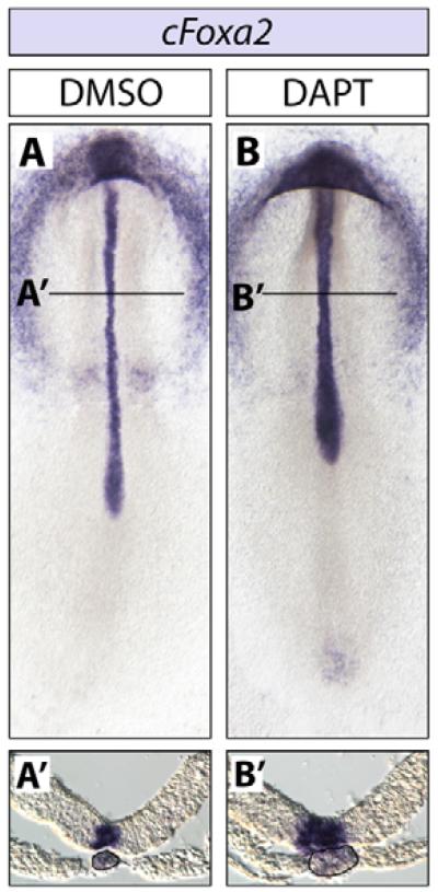
Hensen's node of the chick embryo contains multipotent self-renewing progenitor cells that can contribute to either the floor plate or the notochord. Floor plate cells are a population of epithelial cells that lie at the ventral midline of the developing neural tube, whereas the notochord is a rod of axial mesoderm that lies directly beneath the floor plate. These two tissues serve as a source of a potent signalling morphogen, sonic hedgehog (Shh), which patterns the dorsoventral axis of the neural tube. We show, through both gain- and loss-of-function approaches, that Notch signalling promotes the contribution of chick axial progenitor cells to the floor plate and inhibits contribution to the notochord. Thus, we propose that Notch regulates the allocation of appropriate numbers of progenitor cells from Hensen's node of the chick embryo to the notochord and the floor plate.
Figures
Similar articles
-
A revised model of Xenopus dorsal midline development: differential and separable requirements for Notch and Shh signaling.Dev Biol. 2011 Apr 15;352(2):254-66. doi: 10.1016/j.ydbio.2011.01.021. Epub 2011 Jan 27. Dev Biol. 2011. PMID: 21276789 Free PMC article.
-
Defining subregions of Hensen's node essential for caudalward movement, midline development and cell survival.Development. 1999 Nov;126(21):4771-83. doi: 10.1242/dev.126.21.4771. Development. 1999. PMID: 10518494
-
Dual origin of the floor plate in the avian embryo.Development. 2002 Oct;129(20):4785-96. doi: 10.1242/dev.129.20.4785. Development. 2002. PMID: 12361970
-
[Regression of Hensen's node and axial growth of the embryo].J Soc Biol. 1999;193(3):237-41. J Soc Biol. 1999. PMID: 10542953 Review. French.
-
Neurulation in amniote vertebrates: a novel view deduced from the use of quail-chick chimeras.Int J Dev Biol. 1998;42(7):909-16. Int J Dev Biol. 1998. PMID: 9853821 Review.
Cited by
-
Intrinsic and extrinsic cues time somite progenitor contribution to the vertebrate primary body axis.Elife. 2024 Jan 9;13:e90499. doi: 10.7554/eLife.90499. Elife. 2024. PMID: 38193440 Free PMC article.
-
A revised model of Xenopus dorsal midline development: differential and separable requirements for Notch and Shh signaling.Dev Biol. 2011 Apr 15;352(2):254-66. doi: 10.1016/j.ydbio.2011.01.021. Epub 2011 Jan 27. Dev Biol. 2011. PMID: 21276789 Free PMC article.
-
Isolation and characterization of node/notochord-like cells from mouse embryonic stem cells.Stem Cells Dev. 2011 Nov;20(11):1817-27. doi: 10.1089/scd.2011.0042. Epub 2011 Apr 6. Stem Cells Dev. 2011. PMID: 21351873 Free PMC article.
-
Cellular processes driving gastrulation in the avian embryo.Mech Dev. 2020 Sep;163:103624. doi: 10.1016/j.mod.2020.103624. Epub 2020 Jun 17. Mech Dev. 2020. PMID: 32562871 Free PMC article. Review.
-
ProNodal acts via FGFR3 to govern duration of Shh expression in the prechordal mesoderm.Development. 2015 Nov 15;142(22):3821-32. doi: 10.1242/dev.119628. Epub 2015 Sep 28. Development. 2015. PMID: 26417042 Free PMC article.
References
-
- Amacher SL, Draper BW, Summers BR, Kimmel CB. The zebrafish T-box genes no tail and spadetail are required for development of trunk and tail mesoderm and medial floor plate. Development. 2002;129:3311–3323. - PubMed
-
- Appel B. Zebrafish neural induction and patterning. Dev. Dyn. 2000;219:155–168. - PubMed
-
- Appel B, Fritz A, Westerfield M, Grunwald DJ, Eisen JS, Riley BB. Delta-mediated specification of midline cell fates in zebrafish embryos. Curr. Biol. 1999;9:247–256. - PubMed
-
- Catala M, Teillet MA, Le Douarin NM. Organization and development of the tail bud analyzed with the quail-chick chimaera system. Mech. Dev. 1995;51:51–65. - PubMed
Publication types
MeSH terms
Substances
Grants and funding
LinkOut - more resources
Full Text Sources
