. 2010 Jul 1;87(1):166-76.
doi: 10.1093/cvr/cvq031. Epub 2010 Jan 28.

An angiotensin II- and NF-kappaB-dependent mechanism increases connexin 43 in murine arteries targeted by renin-dependent hypertension

Affiliations

An angiotensin II- and NF-kappaB-dependent mechanism increases connexin 43 in murine arteries targeted by renin-dependent hypertension

Florian Alonso et al. Cardiovasc Res. .

Abstract

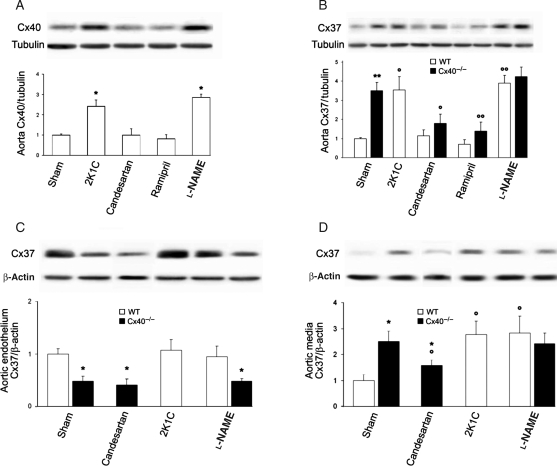
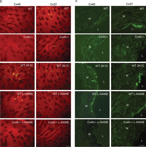
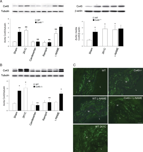
Aims: Connexins (Cxs) play a role in the contractility of the aorta wall. We investigated how connexins of the endothelial cells (ECs; Cx37, Cx40) and smooth muscle cells (SMCs; Cx43, Cx45) of the aorta change during renin-dependent and -independent hypertension.

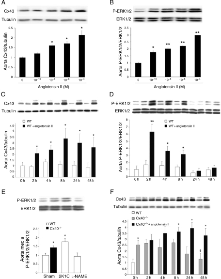
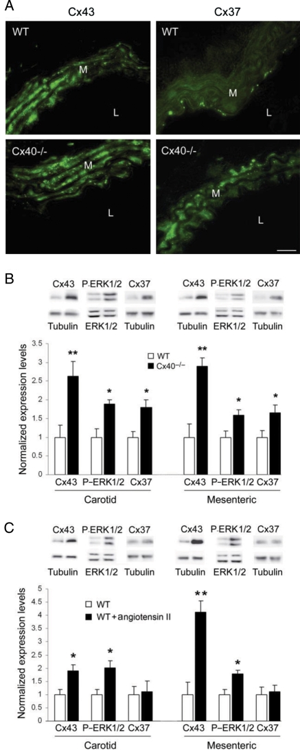
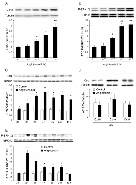
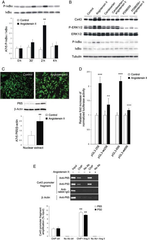
Methods and results: We subjected both wild-type (WT) mice and mice lacking Cx40 (Cx40(-/-)), to either a two-kidney, one-clip procedure or to N-nitro-l-arginine-methyl-ester treatment, which induce renin-dependent and -independent hypertension, respectively. All hypertensive mice featured a thickened aortic wall, increased levels of Cx37 and Cx45 in SMC, and of Cx40 in EC (except in Cx40(-/-) mice). Cx43 was up-regulated, with no effect on its S368 phosphorylation, only in the SMCs of renin-dependent models of hypertension. Blockade of the renin-angiotensin system of Cx40(-/-) mice normalized blood pressure and prevented both aortic thickening and Cx alterations. Ex vivo exposure of WT aortas, carotids, and mesenteric arteries to physiologically relevant levels of angiotensin II (AngII) increased the levels of Cx43, but not of other Cx. In the aortic SMC line of A7r5 cells, AngII activated kinase-dependent pathways and induced binding of the nuclear factor-kappa B (NF-kappaB) to the Cx43 gene promoter, increasing Cx43 expression.

Conclusion: In both large and small arteries, hypertension differently regulates Cx expression in SMC and EC layers. Cx43 is selectively increased in renin-dependent hypertension via an AngII activation of the extracellular signal-regulated kinase and NF-kappaB pathways.


Figures

Figure 1
Figure 1
Cx40 and Cx37 increase in the aorta of hypertensive mice. (A) Western blots showed increased Cx40 in renin-dependent and -independent hypertensive WT mice. (B) Cx37 was also higher in the aortas of hypertensive animals. Treatment with candesartan or ramipril reduced the expression of Cx37 in the spontaneously hypertensive Cx40−/− mice. (C) Western blots demonstrated that Cx37 levels did not change in the aortic intima of either 2K1C or l-NAME hypertensive mice, and were lower in Cx40−/− than in WT animals. Treatment with candesartan did not affect Cx37 levels in the endothelium of Cx40−/− mice. (D) Cx37 was less abundant in the SMCs than in the ECs of WT controls, whereas it increased in the SMCs of Cx40−/− mice. A Cx37 increase occurred in the media of all hypertensive mice. The levels of SMC Cx37 were also altered after candesartan treatment in Cx40−/− mice. Data represent mean + SEM of four experiments. *P < 0.05, **P < 0.01 vs. the respective WT animals; °P < 0.05, °°P < 0.01 vs. WT or Cx40−/− untreated or sham-operated animals. (E) En face immunostaining showed that Cx40, but not Cx37, was increased between ECs of hypertensive mice. (F) Sections confirmed that Cx40 was solely expressed in ECs, whereas Cx37 was detectable in ECs and in the SMCs of hypertensive mice. L, lumen; M, media. Bar represents 30 µm.
Figure 1
Figure 1
Cx40 and Cx37 increase in the aorta of hypertensive mice. (A) Western blots showed increased Cx40 in renin-dependent and -independent hypertensive WT mice. (B) Cx37 was also higher in the aortas of hypertensive animals. Treatment with candesartan or ramipril reduced the expression of Cx37 in the spontaneously hypertensive Cx40−/− mice. (C) Western blots demonstrated that Cx37 levels did not change in the aortic intima of either 2K1C or l-NAME hypertensive mice, and were lower in Cx40−/− than in WT animals. Treatment with candesartan did not affect Cx37 levels in the endothelium of Cx40−/− mice. (D) Cx37 was less abundant in the SMCs than in the ECs of WT controls, whereas it increased in the SMCs of Cx40−/− mice. A Cx37 increase occurred in the media of all hypertensive mice. The levels of SMC Cx37 were also altered after candesartan treatment in Cx40−/− mice. Data represent mean + SEM of four experiments. *P < 0.05, **P < 0.01 vs. the respective WT animals; °P < 0.05, °°P < 0.01 vs. WT or Cx40−/− untreated or sham-operated animals. (E) En face immunostaining showed that Cx40, but not Cx37, was increased between ECs of hypertensive mice. (F) Sections confirmed that Cx40 was solely expressed in ECs, whereas Cx37 was detectable in ECs and in the SMCs of hypertensive mice. L, lumen; M, media. Bar represents 30 µm.
Figure 2
Figure 2
Cx43 and Cx45 are differently affected by renin-dependent and -independent hypertension. (A) Cx45 increased in all WT mice made hypertensive, and this increase was blocked in Cx40−/− mice by RAS inhibitors (left panel). Cx45 was specifically increased in the aortic SMCs of hypertensive mice (right panel). (B) In contrast, Cx43 increased only in aortas of renin-dependent models (Cx40−/− and 2K1C). Data represent mean + SEM of four experiments. *P < 0.05, **P < 0.01 vs. the respective WT animals; °P < 0.05, °°P < 0.01 vs. WT or Cx40−/− untreated or sham-operated animals. (C) Immunostaining showed Cx43 throughout the media of all aortas. Increased numbers of immunospots were observed in the media of all Cx40−/− and hypertensive 2K1C WT mice. M, media; L, lumen. Bar represents 30 µm.
Figure 3
Figure 3
AngII stimulates Cx43 expression in a dose-dependent manner. (A and B) Western blots of WT aortas exposed for 4 h to AngII showed that the peptide increased the levels of Cx43 and P-ERK1/2, at physiologically nanomolar concentrations, and reproduced the two-fold Cx43 increase observed in vivo, at concentrations >0.1 µM. (C and D) Exposure of WT aortas to 2 µM AngII increased within 2 h Cx43 (C), and P-ERK1/2 (D). (E) The levels of the P-ERK1/2 increased in aortas of Cx40−/− and 2K1C mice, but not in those of l-NAME-treated mice. (F) A smaller and delayed effect was observed with aortas of Cx40−/− mice. In the absence of AngII, the levels of Cx43 decreased in this tissue to reach control values within 48 h. Data represent mean + SEM of three experiments. *P < 0.05, **P < 0.01, °P < 0.05 vs. respective untreated WT or Cx40−/− aortas. §P < 0.05 vs. untreated Cx40−/− aortas at t = 0 h.
Figure 4
Figure 4
Carotid and mesenteric arteries show a Cx pattern and responsiveness to AngII similar to those of aorta. (A, left panels) Immunostaining showed punctate Cx43 throughout the carotid media. Increased numbers of immunospots were observed in the media of Cx40−/− mice. (A, right panels) Cx37 was detectable in the media of hypertensive but not control mice. L, lumen; M, media. Bar represents 10 µm. (B) Western blots showed that Cx37, Cx43, and P-ERK1/2 were increased in carotid and mesenteric arteries of Cx40−/− mice. (C) After a 4 h exposure of WT carotids and mesenteric arteries to 2 µM AngII, Cx43 and P-ERK1/2, but not Cx37, were increased. Data represent mean + SEM of three experiments. *P < 0.05, **P < 0.01 vs. respective untreated vessels or WT animals.
Figure 5
Figure 5
A7r5 cells feature the same Cx43 and signalling changes observed in aortic SMCs. (A and B) Western blots of A7r5 cells exposed for 2 h to increasing concentrations of AngII showed that the peptide increased the levels of Cx43 and P-ERK1/2 at concentrations similar to those that elicited comparable changes in aortic SMCs. (C and D) Western blots showed that A7r5 cells expressed Cx43, Cx37, Cx40, and Cx45. Two-micromolar AngII increased Cx43, but not Cx37, Cx40, and Cx45. (E) AngII also increased the levels of P-ERK1/2. Results are means + SEM of three experiments. *P < 0.05, **P < 0.01 vs. untreated cells.
Figure 6
Figure 6
The AngII-induced increase in Cx43 requires the activation of ERK and NF-κB pathways. (A) Within 2 h, AngII increased the phosphorylation of IκBα in A7r5 cells. Results are means + SEM of three experiments. **P < 0.01 vs. untreated cells. (B) Within 2 h, AngII also increased Cx43, P-ERK1/2, and P-IκBα levels. Both candesartan and PD98059 prevented these effects. (C) After 4 h, 2 µM AngII induced a translocation of the P65 phosphorylated form of NF-κB into the nucleus of A7r5 cells. Bar, 20 µm. (D) AngII increased luciferase activity of A7r5 cells transfected with either a short or long Gja1 promoter fragment. Mutation of the NF-κB motif in either one of the two promoter fragments prevented the increased in the luciferase activity observed after AngII stimulation. (E) Quantitative RT–PCR showed that AngII increased the amount of the Gja1 promoter fragment, recovered using antibodies against either the P65 or the P50 forms of NF-κB. Results are means + SEM of three experiments. **P < 0.01, ***P < 0.001 vs. untreated cells; °°P < 0.01, °°°P < 0.001 vs. AngII-treated cells.

Similar articles

Cited by

References

    1. Haefliger JA, Nicod P, Meda P. Contribution of connexins to the function of the vascular wall. Cardiovasc Res. 2004;62:345–356. - PubMed
    1. Simon AM, McWhorter AR. Decreased intercellular dye-transfer and downregulation of non-ablated connexins in aortic endothelium deficient in connexin37 or connexin40. J Cell Sci. 2003;116:2223–2236. - PubMed
    1. Kruger O, Beny JL, Chabaud F, Traub O, Theis M, Brix K, et al. Altered dye diffusion and upregulation of connexin37 in mouse aortic endothelium deficient in connexin40. J Vasc Res. 2002;39:160–172. - PubMed
    1. Brisset AC, Isakson BE, Kwak BR. Connexins in vascular physiology and pathology. Antioxid Redox Signal. 2009;11:267–282. - PMC - PubMed
    1. Haefliger JA, Krattinger N, Martin D, Pedrazzini T, Capponi A, Doring B, et al. Connexin43-dependent mechanism modulates renin secretion and hypertension. J Clin Invest. 2006;116:405–413. - PMC - PubMed

Publication types

MeSH terms