

McConnell taping shifts the patella inferiorly in patients with patellofemoral pain: a dynamic magnetic resonance imaging study
- PMID: 20110340
- PMCID: PMC2836141
- DOI: 10.2522/ptj.20080365
McConnell taping shifts the patella inferiorly in patients with patellofemoral pain: a dynamic magnetic resonance imaging study
Abstract
Background: Patellar taping is widely used clinically to treat patients with patellofemoral pain syndrome (PFPS). Although patellar taping has been demonstrated to reduce patellofemoral pain in patients with PFPS, the kinematic source for this pain reduction has not been identified.
Objective: The purpose of this study was to quantify the changes in the 6-degrees-of-freedom patellofemoral kinematics due to taping in patients with PFPS.
Design: A within-subject design and a sample of convenience were used.
Participants: Fourteen volunteers (19 knees) who were diagnosed with patellofemoral pain that was present for a year or longer were included. Each knee had to meet at least 1 of the following inclusion criteria: Q-angle of > or =15 degrees, a positive apprehension test, patellar lateral hypermobility (> or =10 mm), or a positive "J sign."
Methods: Each knee underwent 2 randomly ordered testing conditions (untaped and taped). A full fast-phase contrast (PC) magnetic resonance image set was acquired for each condition while the participants volitionally extended and flexed their knee. Three-dimensional displacements and rotations were calculated through integration of the fast-PC velocity data. Statistical comparisons between baseline patellofemoral kinematics and the change in kinematics due to taping were performed using a 2-tailed paired Student t test. Correlations between baseline patellofemoral kinematics and the change in kinematics due to taping also were quantified.
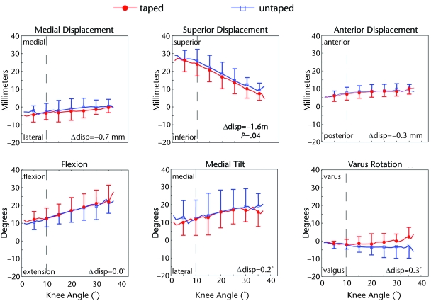
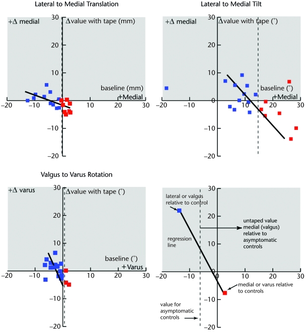
Results: Patellar taping resulted in a significant patellofemoral inferior shift. The strongest correlation existed between the change in lateral-medial displacement with taping and baseline (r=-.60).
Conclusions: The inferior shift in patellar displacement with taping partially explains the previously documented decrease in pain due to increases in contact area. The lack of alteration in 5 of the 6 kinematic variables with taping may have been due to the fact that post-taping kinematic alterations are sensitive to the baseline kinematic values.
Figures
References
-
- Almeida SA, Trone DW, Leone DM, et al. Gender differences in musculoskeletal injury rates: a function of symptom reporting? Med Sci Sports Exerc 1999;31:1807–1812 - PubMed
-
- Rauh MJ, Koepsell TD, Rivara FP, et al. Epidemiology of musculoskeletal injuries among high school cross-country runners. Am J Epidemiol 2006;163:151–159 - PubMed
-
- Cibulka MT, Threlkeld-Watkins J. Patellofemoral pain and asymmetrical hip rotation. Phys Ther 2005;85:1201–1207 - PubMed
-
- Sutlive TG, Mitchell SD, Maxfield SN, et al. Identification of individuals with patellofemoral pain whose symptoms improved after a combined program of foot orthosis use and modified activity: a preliminary investigation. Phys Ther 2004;84:49–61 - PubMed
Publication types
MeSH terms
Substances
Grants and funding
LinkOut - more resources
Full Text Sources
Medical
