. 2010 Mar 15;21(6):1059-71.
doi: 10.1091/mbc.e09-07-0572. Epub 2010 Jan 28.

Assembly of the AAA ATPase Vps4 on ESCRT-III

Affiliations

Assembly of the AAA ATPase Vps4 on ESCRT-III

Anna Shestakova et al. Mol Biol Cell. .

Abstract

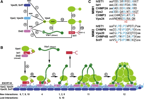
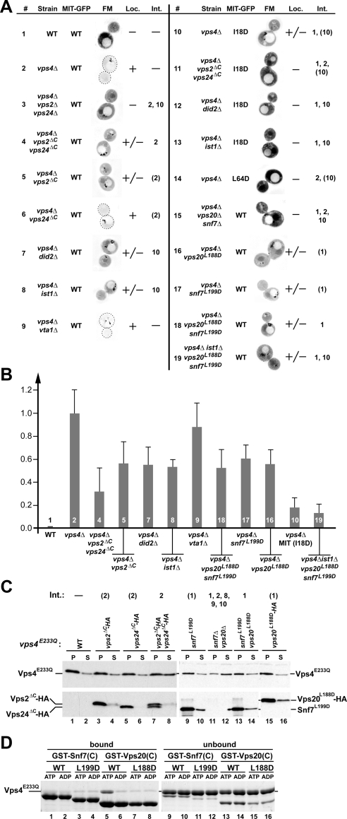
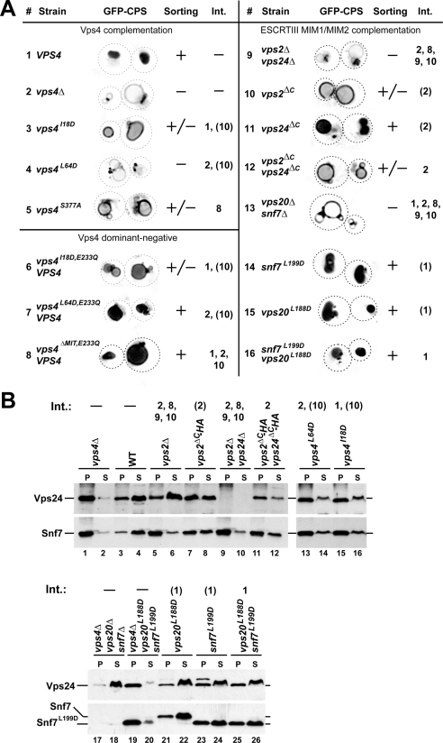
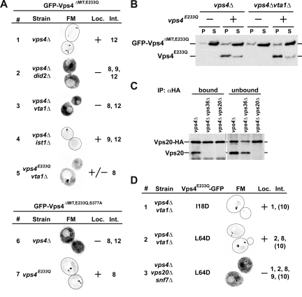
Vps4 is a key enzyme that functions in endosomal protein trafficking, cytokinesis, and retroviral budding. Vps4 activity is regulated by its recruitment from the cytoplasm to ESCRT-III, where the protein oligomerizes into an active ATPase. The recruitment and oligomerization steps are mediated by a complex network of at least 12 distinct interactions between Vps4, ESCRT-III, Ist1, Vta1, and Did2. The order of events leading to active, ESCRT-III-associated Vps4 is poorly understood. In this study we present a systematic in vivo analysis of the Vps4 interaction network. The data demonstrated a high degree of redundancy in the network. Although no single interaction was found to be essential for the localization or activity of Vps4, certain interactions proved more important than others. The most significant among these were the binding of Vps4 to Vta1 and to the ESCRT-III subunits Vps2 and Snf7. In our model we propose the formation of a recruitment complex in the cytoplasm that is composed of Did2-Ist1-Vps4, which upon binding to ESCRT-III recruits Vta1. Vta1 in turn is predicted to cause a rearrangement of the Vps4 interactions that initiates the assembly of the active Vps4 oligomer.


Figures

Figure 1.
Figure 1.
Interactions between Vps4 and its substrate and regulators. (A) Vps4 interaction network based on previous studies (Scott et al., 2005b; Azmi et al., 2008; Kieffer et al., 2008; Bajorek et al., 2009a; Xiao et al., 2009). (B) Model for the recruitment and assembly of Vps4. The numbers indicating New Interactions or Lost Interactions refer to the numbers in A. (C) Alignments of the putative MIM1 and MIM2 motifs of yeast and mammalian ESCRT-III subunits (yeast Ist1 does not contain an obvious MIM2 consensus sequence). Mutations used in this study are marked in red.
Figure 2.
Figure 2.
MIM1 and MIM2 interactions contribute to the recruitment of Vps4 to ESCRT-III. (A) Fluorescence microscopy analysis of the Vps4 MIT domain fused to GFP (MIT-GFP). The wild-type, MIM1 mutant (L64D), or MIM2 mutant (I18D) version of MIT-GFP was expressed in different yeast strains (see Table 1), and the extent of endosomal localization was determined (Loc.). For better visualization the fluorescence microscopy pictures were inverted and the intensity was adjusted to the individual brightness range (black is the brightest signal). Vps4 interactions affected by the different mutations are listed (Int., numbers are based on the interactions in Figure 1A). Numbers in parentheses indicate partially disrupted interactions. (B) Quantification of endosome-localized MIT-GFP relative to wild type (0.0) and vps4Δ (1.0). The data shown represent the results of at least 15 individually analyzed cells. Numbers refer to the experiment number in A. (C) Subcellular fractionation of different yeast strains expressing vps4E233Q into soluble, cytoplasmic fraction (S) and pelletable, membrane-associated fraction (P). Fractions were analyzed by Western blot using antibodies specific for Vps4 (top panels), Snf7 (bottom panel, lanes 9–14) and the HA-tag (bottom panels, lanes 1–8 and 15–16). Vps4 interactions affected by the different mutations are listed (Int., numbers are based on the interactions in Figure 1A). Numbers in parentheses indicate partially disrupted interactions. (D) In vitro Vps4 interaction studies using wild-type and MIM2 mutant forms of GST-Vps20(C) (fusion of GST with C-terminal half of Vps20) or GST-Snf7(C) immobilized on GSH-Sepharose. Vps4E233Q was added in the presence of ATP or ADP to immobilized proteins; bound and unbound fractions were analyzed by SDS-PAGE and Coomassie staining.
Figure 3.
Figure 3.
Phenotypic analysis of mutations affecting Vps4 interactions. (A) Fluorescence microscopy analysis of yeast strains expressing GFP-CPS. The efficiency of GFP-CPS sorting into the lumen of the vacuole is indicated (+, +/−, −). Vps4 interactions affected by the different mutations are listed. (B) Subcellular fractionation of yeast strains into soluble (S) and membrane-associated pellet fractions (P). The samples were analyzed by Western blot using antibodies specific for Vps24 and Snf7. Vps4 interactions affected by the different mutations are listed. (A and B) Int., numbers are based on the interactions in Figure 1A, and numbers in parentheses indicate partially disrupted interactions.
Figure 4.
Figure 4.
Localization of mutant Vps4 proteins. (A) Localization of MIT-deleted Vps4 (GFP- Vps4ΔMIT,E233Q and GFP-Vps4ΔMIT,E233Q,S377A) in different yeast mutant strains (see Table 1) determined by fluorescence microscopy (FM). (A and D) The extent of observed endosomal localization is indicated (Loc.). Vps4 interactions affected by the different mutations are listed. Int., numbers are based on the interactions in Figure 1A. (B) Endosomal recruitment of MIT-deleted Vps4 in the presence or absence of full-length Vps4 protein determined by subcellular fractionation and Western blot analysis (S, soluble; P, pellet). (C) Immunoprecipitation of Vps20-HA from detergent-solubilized membrane fractions. The resulting bound and unbound samples were analyzed by Western blot using anti-Vps20 antiserum. (D) Fluorescence microscopy (FM) analysis of GFP-tagged full-length Vps4 protein in different mutant strains (Table 1). Numbers in parentheses indicate partially disrupted interactions.
Figure 5.
Figure 5.
Ist1 and Did2 form a stable complex. GST-tagged full-length Did2 (GST-Did2) or C-terminal half of Did2 [GST-CT(Did2)] was immobilized on GSH-Sepharose, and purified Ist1, Vta1, or Vps4E233Q protein was added. The resulting bound and unbound fractions were analyzed by SDS-PAGE and Coomassie staining.

References

    1. Agromayor M., Carlton J. G., Phelan J. P., Matthews D. R., Carlin L. M., Ameer-Beg S., Bowers K., Martin-Serrano J. Essential role of hIST1 in cytokinesis. Mol. Biol. Cell. 2009;20:1374–1387. - PMC - PubMed
    1. Azmi I., Davies B., Dimaano C., Payne J., Eckert D., Babst M., Katzmann D. J. Recycling of ESCRTs by the AAA-ATPase Vps4 is regulated by a conserved VSL region in Vta1. J. Cell Biol. 2006;172:705–717. - PMC - PubMed
    1. Azmi I. F., Davies B. A., Xiao J., Babst M., Xu Z., Katzmann D. J. ESCRT-III family members stimulate Vps4 ATPase activity directly or via Vta1. Dev Cell. 2008;14:50–61. - PubMed
    1. Babst M. A protein's final ESCRT. Traffic. 2005;6:2–9. - PubMed
    1. Babst M., Katzmann D. J., Estepa-Sabal E. J., Meerloo T., Emr S. D. Escrt-III: an endosome-associated heterooligomeric protein complex required for mvb sorting. Dev. Cell. 2002;3:271–282. - PubMed

Publication types

MeSH terms

Substances

LinkOut - more resources