

Dose exposure of patients undergoing comprehensive stroke imaging by multidetector-row CT: comparison of 320-detector row and 64-detector row CT scanners
- PMID: 20110373
- PMCID: PMC7963949
- DOI: 10.3174/ajnr.A1971
Dose exposure of patients undergoing comprehensive stroke imaging by multidetector-row CT: comparison of 320-detector row and 64-detector row CT scanners
Abstract
Background and purpose: Recently introduced 320-detector row CT enables whole brain perfusion imaging compared to a limited scanning area in 64-detector row CT. Our aim was to evaluate patient radiation exposure in comprehensive stroke imaging by using multidetector row CT consisting of standard CT of the head, CTA of cerebral and cervical vessels, and CTP.
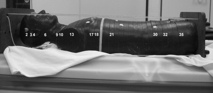
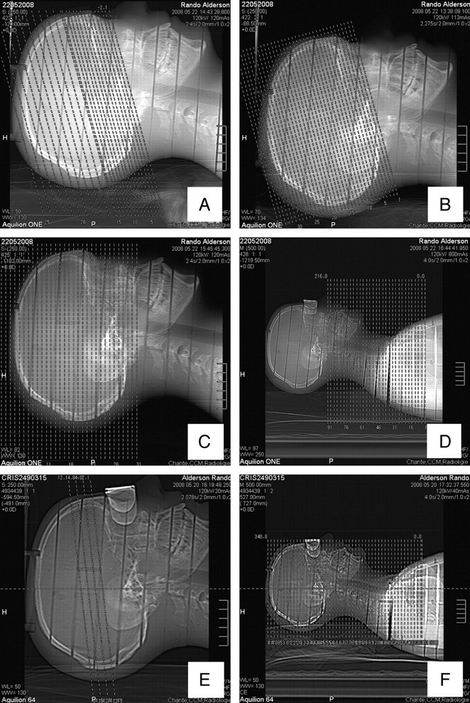
Material and methods: Organ doses were measured by using LiF-TLDs located at several organ sites in an Alderson-Rando phantom. Effective doses were derived from these measurements. Stroke protocols including noncontrast head CT, CTA of cerebral and cervical vessels, and CTP were performed on 320- and 64-detector row scanners.
Results: Measured effective doses for the different scanning protocols ranged between 1.61 and 4.56 mSv, resulting in an effective dose for complete stroke imaging of 7.52/7.54 mSv (m/f) for 64-detector row CT and 10.56/10.6 mSv (m/f) for 320-detector row CT. The highest organ doses within the area of the primary beam were measured in the skin (92 mGy) and cerebral hemispheres (69.91 mGy). Use of an eye-protection device resulted in a 54% decrease of the lens dose measured for the combo protocol for whole-brain perfusion with the 320-detector row CT scanner.
Conclusions: Phantom measurements indicate that comprehensive stroke imaging with multidetector row CT may result in effective radiation doses from 7.52 mSv (64-detector row CT) to 10.6 mSv (320-detector row CT). The technique of 320-detector row CT offers additional information on the time course of vascular enhancement and whole-brain perfusion. Physicians should weigh the potential of the new technique against the higher radiation dose that is needed. Critical doses that would cause organ damage were not reached.
Figures
Similar articles
-
Radiation dose evaluation in multidetector-row CT imaging for acute stroke with an anthropomorphic phantom.Br J Radiol. 2010 Dec;83(996):1029-41. doi: 10.1259/bjr/52267127. Br J Radiol. 2010. PMID: 21088088 Free PMC article.
-
Effective dose to patient measurements for flat-detector computed tomography protocols in acute stroke care.Eur Radiol. 2020 Sep;30(9):5082-5088. doi: 10.1007/s00330-020-06891-w. Epub 2020 Apr 28. Eur Radiol. 2020. PMID: 32346793
-
Radiation exposure of patients in comprehensive computed tomography of the head in acute stroke.AJNR Am J Neuroradiol. 2006 Sep;27(8):1741-5. AJNR Am J Neuroradiol. 2006. PMID: 16971627 Free PMC article.
-
Area-Detector Computed Tomography for Pulmonary Functional Imaging.Diagnostics (Basel). 2023 Jul 28;13(15):2518. doi: 10.3390/diagnostics13152518. Diagnostics (Basel). 2023. PMID: 37568881 Free PMC article. Review.
-
State of the art: technologies for computed tomography dose reduction.Emerg Radiol. 2010 May;17(3):209-18. doi: 10.1007/s10140-009-0850-6. Epub 2009 Nov 20. Emerg Radiol. 2010. PMID: 19936808 Review.
Cited by
-
Case series of 64 slice computed tomography-computed tomographic angiography with 3D reconstruction to diagnose symptomatic cerebral aneurysms: new standard of care?Neurol Int. 2012 Jan 9;4(1):e2. doi: 10.4081/ni.2012.e2. Epub 2012 Feb 23. Neurol Int. 2012. PMID: 22593806 Free PMC article.
-
Cut-Out Towne-View Whole-Brain 320-Row Four-Dimensional Computed Tomography Angiography for Assessing the Anterior Intracranial Collateral Status: A Retrospective Study.Diagnostics (Basel). 2022 May 27;12(6):1336. doi: 10.3390/diagnostics12061336. Diagnostics (Basel). 2022. PMID: 35741146 Free PMC article.
-
KERMA ratios in pediatric CT dosimetry.Pediatr Radiol. 2012 May;42(5):527-35. doi: 10.1007/s00247-011-2336-4. Epub 2012 Mar 20. Pediatr Radiol. 2012. PMID: 22430480
-
CT-perfusion in peripheral arterial disease - Correlation with angiographic and hemodynamic parameters.PLoS One. 2019 Sep 27;14(9):e0223066. doi: 10.1371/journal.pone.0223066. eCollection 2019. PLoS One. 2019. PMID: 31560706 Free PMC article.
-
Estimation of Absorbed Dose of the Thyroid Gland in Patients Undergoing 64-Slice Head Computed Tomography and Comparison the Results with ImPACT Software and Computed Tomography Scan Dose Index.J Med Signals Sens. 2019 Aug 29;9(3):190-195. doi: 10.4103/jmss.JMSS_40_18. eCollection 2019 Jul-Sep. J Med Signals Sens. 2019. PMID: 31544059 Free PMC article.
References
-
- Bohner G, Förschler A, Hamm B, et al. . Quantitative perfusion imaging by multi-slice CT in stroke patients [in German]. Rofo 2003;1785:806–13 - PubMed
-
- Hoeffner E, Case I, Jain R, et al. . Cerebral perfusion CT: technique and clinical applications. Radiology 2004;231:632–44. Epub 2004 Apr 29 - PubMed
-
- Boone JM. The trouble with CTDI 100. Med Phys 2007;34:1364–71 - PubMed
-
- Kyriakou Y, Deak P, Langner O, et al. . Concepts for dose determination in flat-detector CT. Phys Med Biol 2008;53:3551–66. Epub 2008 Jun 13 - PubMed
MeSH terms
LinkOut - more resources
Full Text Sources
Medical