

Controlled Clinical Trial
doi: 10.3174/ajnr.A2008.
Epub 2010 Jan 28.
Multiple inflow pulsed arterial spin-labeling reveals delays in the arterial arrival time in minor stroke and transient ischemic attack
Affiliations
- PMID: 20110375
- PMCID: PMC7964001
- DOI: 10.3174/ajnr.A2008
Item in Clipboard
Controlled Clinical Trial
Multiple inflow pulsed arterial spin-labeling reveals delays in the arterial arrival time in minor stroke and transient ischemic attack
AJNR Am J Neuroradiol.
2010 Nov.
Abstract
Our purpose was to use multiple inflow pulsed ASL to investigate whether hemodynamic AAT information is sensitive to hemispheric asymmetry in acute ischemia. The cohorts included 15 patients with acute minor stroke or TIA and 15 age-matched controls. Patients were scanned by using a stroke MR imaging protocol at a median time of 74 hours. DWI lesion volumes were small and functional impairment was low; however, perfusion abnormalities were evident. Prolonged AAT values were more likely to reside in the affected hemisphere (significant when compared with controls, P < .048). An advantage of this ASL technique is the ability to use AAT information in addition to CBF to characterize ischemia.
Figures

Acute images for 3 patients with decreasing infarct volumes. Patient 1 shows a delayed AAT region (hyperintensity) in the affected hemisphere, where CBF is reduced (hypointensity). Patient 12 shows reduced CBF in the affected hemisphere and delayed AAT, while maps for Patient 9 have normal findings. Abnormal CBF and AAT regions are outlined in red.
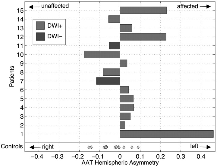
AAT hemispheric asymmetry index is displayed as a bar plot for each patient. Control cohort data are more symmetric about zero with less variance. Patient data are significantly different from those of controls (P < .048). Light gray indicates DWI-positive; dark gray, DWI-negative; diamond datum, control participant.
References
-
- Detre JA, Alsop DC, Vives LR, et al. Noninvasive MRI evaluation of cerebral blood flow in cerebrovascular disease. Neurology 1998;50:633–41 - PubMed
-
- Chalela JA, Alsop DC, Gonzalez-Atavales JB, et al. Magnetic resonance perfusion imaging in acute ischemic stroke using continuous arterial spin labeling. Stroke 2000;31:680–87 - PubMed
-
- Hendrikse J, van Osch MJ, Rutgers DR, et al. Internal carotid artery occlusion assessed at pulsed arterial spin-labeling perfusion MR imaging at multiple delay times. Radiology 2004;233:899–904. Epub 2004 Oct 14 - PubMed
-
- van Laar PJ, Hendrikse J, Klijn CJ, et al. Symptomatic carotid artery occlusion: flow territories of major brain-feeding arteries. Radiology 2007;242:526–34 - PubMed
Publication types
MeSH terms
Substances
Grants and funding
LinkOut - more resources
Full Text Sources
Other Literature Sources
Medical
Research Materials
Miscellaneous