. 2010 Mar;176(3):1130-8.
doi: 10.2353/ajpath.2010.090832. Epub 2010 Jan 28.

Hypoxia inducible-factor1alpha regulates the metabolic shift of pulmonary hypertensive endothelial cells

Affiliations

Hypoxia inducible-factor1alpha regulates the metabolic shift of pulmonary hypertensive endothelial cells

Iwona Fijalkowska et al. Am J Pathol. 2010 Mar.

Abstract

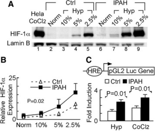
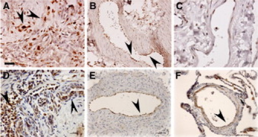
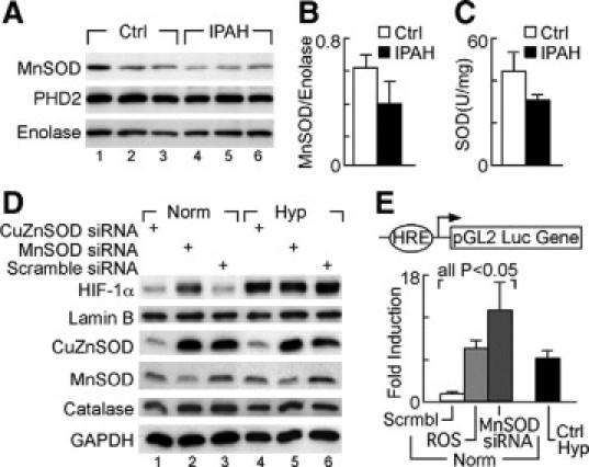
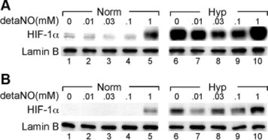
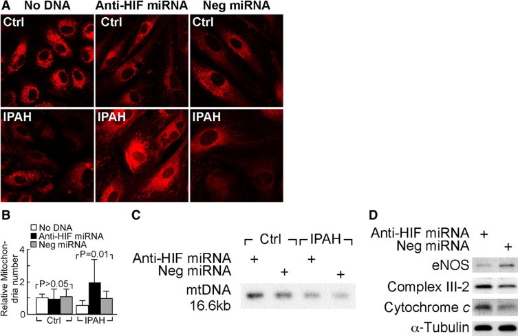
Severe pulmonary hypertension is irreversible and often fatal. Abnormal proliferation and resistance to apoptosis of endothelial cells (ECs) and hypertrophy of smooth muscle cells in this disease are linked to decreased mitochondria and preferential energy generation by glycolysis. We hypothesized this metabolic shift of pulmonary hypertensive ECs is due to greater hypoxia inducible-factor1alpha (HIF-1alpha) expression caused by low levels of nitric oxide combined with low superoxide dismutase activity. We show that cultured ECs from patients with idiopathic pulmonary arterial hypertension (IPAH-ECs) have greater HIF-1alpha expression and transcriptional activity than controls under normoxia or hypoxia, and pulmonary arteries from affected patients have increased expression of HIF-1alpha and its target carbonic anhydrase IX. Decreased expression of manganese superoxide dismutase (MnSOD) in IPAH-ECs paralleled increased HIF-1alpha levels and small interfering (SI) RNA knockdown of MnSOD, but not of the copper-zinc SOD, increased HIF-1 protein expression and hypoxia response element (HRE)-driven luciferase activity in normoxic ECs. MnSOD siRNA also reduced nitric oxide production in supernatants of IPAH-ECs. Conversely, low levels of a nitric oxide donor reduced HIF-1alpha expression in normoxic IPAH-ECs. Finally, mitochondria numbers increased in IPAH-ECs with knockdown of HIF-1alpha. These findings indicate that alterations of nitric oxide and MnSOD contribute to pathological HIF-1alpha expression and account for lower numbers of mitochondria in IPAH-ECs.


Figures

Figure 1
Figure 1
Enhanced HIF-1α expression and transcriptional activity in IPAH-ECs. A: Representative Western blot analysis showing HIF-1α expression in nuclear extracts of IPAH-ECs (n = 3) and control cells (Ctrl, n = 3) under normoxia (Norm) or under a range of hypoxia (Hyp) for 4 hours. B: Quantitative densitometric analysis of Western blots for HIF-1α expression in nuclear extracts of IPAH-ECs (n = 3) and control ECs (n = 3). C: HIF-1α-luciferase reporter activity in cells, transiently transfected with wild-type or mutant HRE-luciferase reporter and cotransfected with renilla construct 24 hours after being exposed to 2.5% O2 hypoxia, treated with 125 μmol/L CoCl2, or left untreated under normoxia. Data shown as fold induction over normoxia expression levels (n ≥4 replicate experiments).
Figure 2
Figure 2
Expression of HIF-1α and its transcriptional target CAIX in IPAH plexiform lesions and remodeled pulmonary arteries. Note increased expression of HIF-1α in cells forming the vascular slits in IPAH plexiform lesions (A, arrowheads) and intimal cells lining the remodeled pulmonary arteries (B, arrowheads), when compared with intima of a normal pulmonary artery (C). This pattern of HIF-1α expression correlated with expression of its transcriptional target CAIX in an IPAH plexiform lesion (D, arrowheads) and pulmonary artery intima (E, arrowhead) when compared with a normal pulmonary artery (F, arrowhead). Scale bars: 25 μm (A, D, E); 50 μm (B, C, F). Images representative of overall five IPAH and five normal lungs tested.
Figure 3
Figure 3
Reduction of MnSOD and SOD activity in IPAH-ECs and regulation of HIF-1α expression by MnSOD in HUVECs. AB: Western blot analysis of MnSOD and prolyl hydroxylase 2 protein expression in whole cell extracts of IPAH (n = 5) and control pulmonary artery ECs (n = 3), normalized by enolase expression (one-way analysis of variance, P = 0.0374). C: SOD activity (U/mg) is reduced in pulmonary arterial hypertension (n = 5), as compared with control pulmonary artery ECs (n = 3) (one way analysis of variance, P = 0.0275). D: HIF-1α expression measured by Western blot of nuclear extracts of HUVECs transiently transfected with MnSOD siRNA, CuZnSOD, or scramble siRNA exposed to 2.5% O2 (Hyp) or normoxia for 4 hours, normalized for nuclear lamin B. MnSOD, CuZnSOD, and catalase protein expression were evaluated in cytosolic fractions, and normalized for cytosolic glyceraldehyde phosphate dehydrogenase. E: HRE activity in HUVECs transiently transfected with wild-type HRE-luciferase reporter and renilla, and either cotransfected with MnSOD siRNA or scramble siRNA (Scrmbl), or treated with pyrogallol (ROS) for 24 hours under normoxia (Norm). Positive controls consisted of HUVECs exposed to 2.5% O2 for 24 hours (Ctrl Hyp).
Figure 4
Figure 4
Nitric oxide modulates HIF-1α expression in IPAH-ECs. IPAH (A) and control (B) pulmonary artery ECs were treated with the NO donor detaNO (10 μmol/L, 30 μmol/L, 100 μmol/L, and 1 mmol/L) or vehicle under normoxia (Norm) or 2.5% O2 (Hyp). HIF-1α expression was determined in nuclear extracts, normalized for expression of lamin-B.
Figure 5
Figure 5
Knockdown HIF-1α by miRNA increases mitochondria in IPAH-pulmonary artery ECs. A and B: Mitochondria were quantified with Mitotracker Red 580 followed by confocal laser-scanning microscopy in IPAH and control pulmonary artery ECs transiently transfected with anti HIF-1α miRNA, negative control miRNA (Neg miRNA), or left untransfected (No DNA). C: Southern analysis of mtDNA content in representative IPAH-ECs and control endothelial cells transfected with anti-HIF-1α miRNA or scrambled miRNA. D: Expression of eNOS, mitochondrial complex III-2, and cytochrome c protein of representative IPAH-EC and control endothelial cells transfected with anti-HIF-1α miRNA or scrambled miRNA assessed by Western blot analysis.

References

    1. Lee SD, Shroyer KR, Markham NE, Cool CD, Voelkel NF, Tuder RM. Monoclonal endothelial cell proliferation is present in primary but not secondary pulmonary hypertension. J Clin Invest. 1998;101:927–934. - PMC - PubMed
    1. Deng Z, Morse JH, Slager SL, Cuervo N, Moore KJ, Venetos G, Kalachikov S, Cayanis E, Fischer SG, Barst RJ, Hodge SE, Knowles JA. Familial primary pulmonary hypertension (gene PPH1) is caused by mutations in the bone morphogenetic protein receptor-II gene. Am J Hum Genet. 2000;67:737–744. - PMC - PubMed
    1. The International PPH Consortium, Lane KB, Machado RD, Pauciulo. M.W., Thompson JR, Philips JA, III, Loyd JE, Nichols WC, Trembath RC. Heterozygous germline mutations in BMPR2 encoding a TGF-B receptor cause familiar pulmonary hypertension. Nat Genet. 2000;26:81–84. - PubMed
    1. Voelkel NF, Cool CD, Lee SD, Wright L, Geraci MW, Tuder RM. Primary pulmonary hypertension between inflammation and cancer. Chest. 1999;114:225S–230S. - PubMed
    1. Rai PR, Cool CD, King JAC, Stevens T, Burns N, Winn RA, Kasper M, Voelkel NF. The cancer paradigm of severe pulmonary arterial hypertension. Am J Respir Crit Care Med. 2008;178:558–564. - PMC - PubMed

Publication types

MeSH terms