. 2010 Mar;176(3):1377-89.
doi: 10.2353/ajpath.2010.090849. Epub 2010 Jan 28.

Pathology of gastrointestinal organs in a porcine model of cystic fibrosis

Affiliations

Pathology of gastrointestinal organs in a porcine model of cystic fibrosis

David K Meyerholz et al. Am J Pathol. 2010 Mar.

Abstract

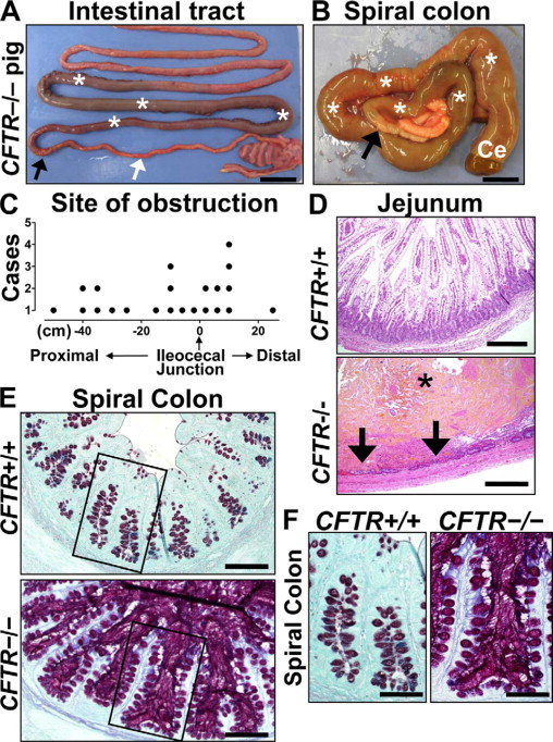
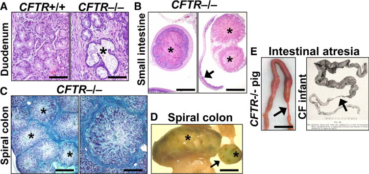
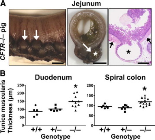
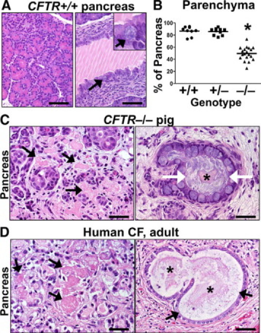
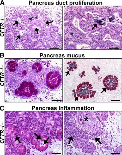
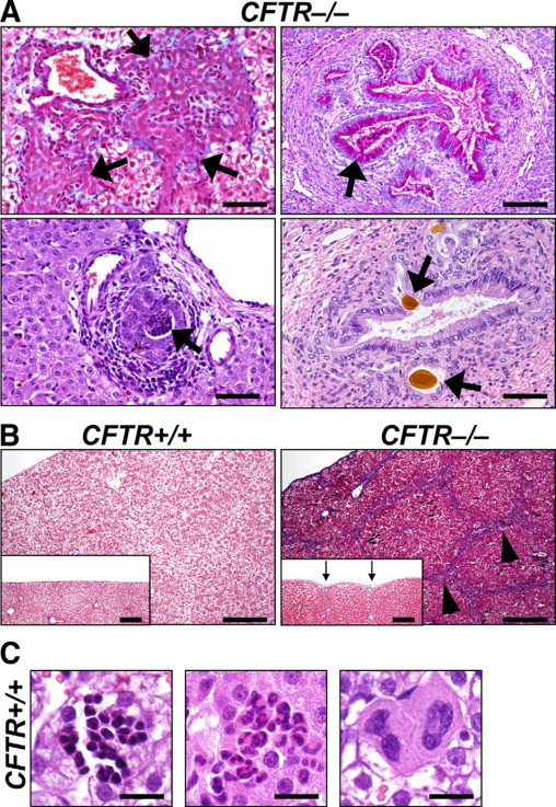
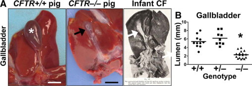
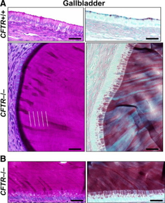
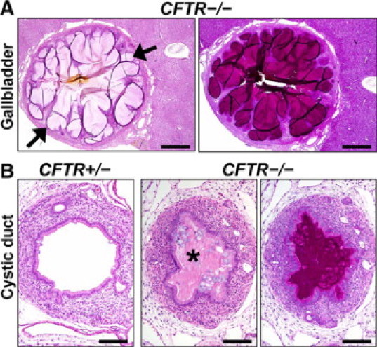
Cystic fibrosis (CF), which is caused by mutations in the gene encoding the cystic fibrosis transmembrane conductance regulator (CFTR), is characterized by multiorgan pathology that begins early in life. To better understand the initial stages of disease, we studied the gastrointestinal pathology of CFTR-/- pigs. By studying newborns, we avoided secondary changes attributable to environmental interactions, infection, or disease progression. Lesions resembling those in humans with CF were detected in intestine, pancreas, liver, gallbladder, and cystic duct. These organs had four common features. First, disease was accelerated compared with that in humans, which could provide a strategy to discover modifying factors. Second, affected organs showed variable hyperplastic, metaplastic, and connective tissue changes, indicating that remodeling was a dynamic component of fetal life. Third, cellular inflammation was often mild to moderate and not always present, which raises new questions as to the role of cellular inflammation in early disease pathogenesis. Fourth, epithelial mucus-producing cells were often increased, producing a striking accumulation of mucus with a layered appearance and resilient structure. Thus, mucus cell hyperplasia and mucus accumulation play prominent roles in early disease. Our findings also have implications for CF lung disease, and they lay the foundation for a better understanding of CF pathogenesis.


Figures

Figure 1
Figure 1
Meconium ileus in CFTR−/− intestine. A: Meconium-filled bowel (asterisks) was proximal to the obstruction interface (black arrow); distal to the interface, the bowel was hypoplastic (white arrow). Scale bar = 2.45 cm. B: Meconium (white asterisks) dilated the spiral colon (ventral view) and cecum (Ce) proximal to the obstruction interface (black arrow). Microcolon appeared distally. Scale bar = 1.34 cm. C: Sites of obstruction in CFTR−/− pigs were distributed on either side of the ileocecal junction extending from the small intestine (proximal) into the spiral colon (distal). D:CFTR−/− jejunum showing luminal meconium (asterisk) causing severe distension and mucosal thinning with focal hemorrhage, necrosis (arrows), and neutrophilic inflammation, HE stain. Scale bars = 0.29 mm. E:CFTR−/− spiral colon had luminal mucus accumulation with hyperplastic mucus-producing cells especially noticeable compared with CFTR+/+ along the surface epithelium (inset boxes, see F). ABPY stain. Scale bars = 117 μm. F:CFTR−/− spiral colon glands were distended by stringy mucus that extends from stout mucus-producing cells in the epithelium, ABPY stain. Scale bars = 69 μm.
Figure 2
Figure 2
Mucus accumulation and atresia in CFTR−/− intestine. A: Duodenal Brünner glands were focally distended by stringy mucus (asterisk) with flattening of adjacent epithelium. HE stain. Scale bars = 80 μm. B: Distal to the obstruction interface, mucocellular cords (asterisks) filled the CFTR−/− intestinal lumen (left panel) and sometimes became dislodged from the intestinal wall to aggregate (right panel) distally in the bowel (arrow). HE stain. Scale bars = 1.2 mm. C: Cross section of a distended CFTR−/− spiral colon filled with large aggregates of mucocellular cords (asterisks) that retained their morphology from the small intestine (see also B, left panel). ABPY stain. Scale bars = 1.7 mm (left panel) and 0.7 mm (right panel). D: Aggregates of mucocellular cords (asterisks) distended a loop of CFTR−/− spiral colon and were detectable in cross section (arrow). Scale bar = 11.1 mm. E: Segmental atresia (arrows) was present distal to the obstruction interface in the CFTR−/− pig (Scale bar = 11.5 mm) and is similar to what Bodian described in CF infants. Reprinted from Fibrocystic Disease of the Pancreas: a Congenital Disorder of Mucus Production-Mucosis, Martin Bodian, Page 84, Copyright 1953.
Figure 3
Figure 3
Diverticuli and smooth muscle thickening in CFTR−/− intestine. A: Diverticula (asterisks and white arrows) formed along the mesenteric border and were lined by a thin mucosa and submucosa, which herniated through a thickened tunica muscularis (black arrows). Scale bars = 3.8 mm (left and middle panel) and 1.4 mm (right panel, HE stain). B: Tunica muscularis of the CFTR−/− pig intestine was generally hypertrophied, including in the duodenum (left, P < 0.01 versus CFTR+/+, P < 0.05, versus CFTR+/−) and the spiral colon (right, P < 0.01 versus CFTR+/+, P < 0.001 versus CFTR+/−, one-way analysis of variance with Bonferroni post test).
Figure 4
Figure 4
Exocrine pancreatic destruction in CFTR−/− pigs. A:CFTR+/+ pig pancreas was rich in exocrine tissue (left panel) with large ducts (right panel) that were partially filled with normal zymogen secretions (white lines are sectioning artifact). Mucus cells were uncommon in the epithelium (arrows and inset). HE stain. Scale bars = 72 μm. B:CFTR−/− pancreata had reduced lobular parenchyma (P < 0.001 vs. CFTR+/+ and CFTR+/−, one-way analysis of variance with Bonferroni post test). Scale bar = mean. C: Degenerative pools of eosinophilic zymogen secretions were detected “free” within the interstitium (black arrows). Within dilated ducts, centrally oriented zymogen secretions (asterisk) were often surrounded by stringy mucus (white arrows). HE stain. Scale bars = 37 μm. D: Adult human CF pancreas (with autolysis making some features less appreciable) showed similar degenerative pools of interstitial zymogen secretions (arrows, left panel) and similar secretions (asterisk) were detected in dilated ducts surrounded by mucus (arrows, right panel). HE stain. Scale bars = 35 μm and 53 μm (left and right panel, respectively).
Figure 5
Figure 5
Duct proliferation, mucus accumulation, and inflammation in CFTR−/− pancreas. A: Foci of proliferative (left panel, arrows) and dilated ducts were common in CFTR−/− pancreata. Dystrophic calcification was rarely detected (right panel, arrow). HE stain. Scale bars = 110 μm and 55 μm (left and right panel, respectively). B: In severely disease pancreata, increased mucinous metaplasia of ducts was detected. Note the large mucus cells (arrows) circumferentially lining variably sized ducts that are moderately distended by stringy mucus. PAS (left panel) and ABPY (right panel) stains. Scale bars = 55 μm. C: Acini and ducts dilated by zymogen material had scattered infiltrates of neutrophils (arrows, left panel) and to a lesser extent, macrophages. Severe exocrine destruction was often associated with interstitial lymphoid aggregates (arrows, right panel) adjacent to dilated, cyst-like ducts (black asterisk). HE stain. Scale bars = 36 μm (left panel) and 55 μm (right panel).
Figure 6
Figure 6
Rare lesions in CFTR−/− liver and incidental findings in all genotypes. A: Infrequent to rare findings in CFTR−/− liver. Biliary tracts had expansive and florid proliferation (arrow, top left panel, MT stain) with adjacent fibrosis (blue) and inflammation. Intrahepatic ducts had mucinous change (arrow, top right panel, PAS stain), mucocellular plugs (arrow, left bottom panel, HE stain), or choleliths (arrows, right bottom panel, HE stain). Scale bars = 55 μm. B: Widespread bridging (arrowheads) of triads by biliary hyperplasia, inflammation, and fibrosis (blue staining) was rarely detected in CFTR−/− pigs. Scale bars = 220 μm. In these cases, the serosal surface was often retracted causing a slightly irregular surface (arrows, inset, Scale bars = 197 μm). MT stain. C: Foci of extramedullary hematopoiesis were composed of erythroid, granulocytic, and megakaryocytic lineage aggregates (respective panels) in liver of all genotypes. HE stain. Scale bars = 17.5 μm.
Figure 7
Figure 7
Microgallbladder in CFTR−/− pigs. A:CFTR+/+ pig gallbladders (asterisk, Scale bar = 10.5 mm) were typically distended with bile, whereas microgallbladder (arrows, Scale bar = 7.4 mm) was seen in the CFTR−/− pig. Right panel shows microgallbladder from infant with CF as reported by Bodian. Reprinted from Fibrocystic Disease of the Pancreas: a Congenital Disorder of Mucus Production-Mucosis, Martin Bodian, Page 112, Copyright 1953. B: Histological measurements showed that porcine CFTR−/− gallbladder was smaller than CFTR+/+ or CFTR+/− (P < 0.001 respectively, one-way analysis of variance with Bonferroni post test, Scale bar = mean).
Figure 8
Figure 8
Mucus cell proliferation and mucus accumulation in CFTR−/− gallbladder. A: Nominal apical mucus staining in CFTR+/+ gallbladder epithelium (top panels, Scale bars = 38 μm). CFTR−/− gallbladder typically had diffuse mucinous change (bottom panels, Scale bars = 26 μm) with contrasting morphological characteristics of mucus including lamellar striations (see white dotted lines) parallel to the epithelium (left panel, PAS stain) and ribbons of resilient mucus perpendicular to the epithelium (right panel, ABPY stain). B: Serial sections of CFTR−/− gallbladder contained similar morphological changes. Scale bars = 32 μm.
Figure 9
Figure 9
Gallbladder and cystic duct lesions. A: The CFTR−/− gallbladder mucosa sometimes formed folds of proliferative epithelium to form cyst-like structures (arrows) along the mucosa. HE (left) and PAS (right) stain. Scale bars = 550 μm. B:CFTR−/− cystic ducts were variably obstructed (asterisk) and stenotic compared with controls. HE (left and middle) and PAS (right) stains. Scale bars = 116 μm.

Similar articles

Cited by

References

    1. Landsteiner K. Darmverschluss durch eingedictes Meconium Pankreatitis. Zentralbl Allg Pathol. 1905;16:903–907.
    1. Andersen DH. Cystic fibrosis of the pancreas and its relation to celiac disease: a clinical and pathologic study. Am J Dis Child. 1938;56:344–399.
    1. Farber S. Pancreatic function and disease early in life V. Pathologic changes associated with pancreatic insufficiency in early life. Arch Path. 1944;37:238–250.
    1. Farber S. Some organic digestive disturbances early in life. J Mich Med Soc. 1946;44:587–594.
    1. Bodian M. Fibrocystic Disease of the Pancreas: a Congenital Disorder of Mucus Production-Mucosis. Grune & Stratton; New York, NY: 1953. pp. 67–146.

Publication types

MeSH terms

Substances