. 2010 Feb 19;327(5968):973-7.
doi: 10.1126/science.1183147. Epub 2010 Jan 28.

N-terminal acetylation of cellular proteins creates specific degradation signals

Affiliations

N-terminal acetylation of cellular proteins creates specific degradation signals

Cheol-Sang Hwang et al. Science. .

Abstract

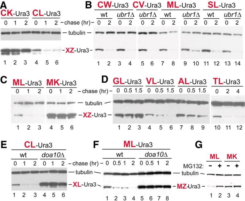
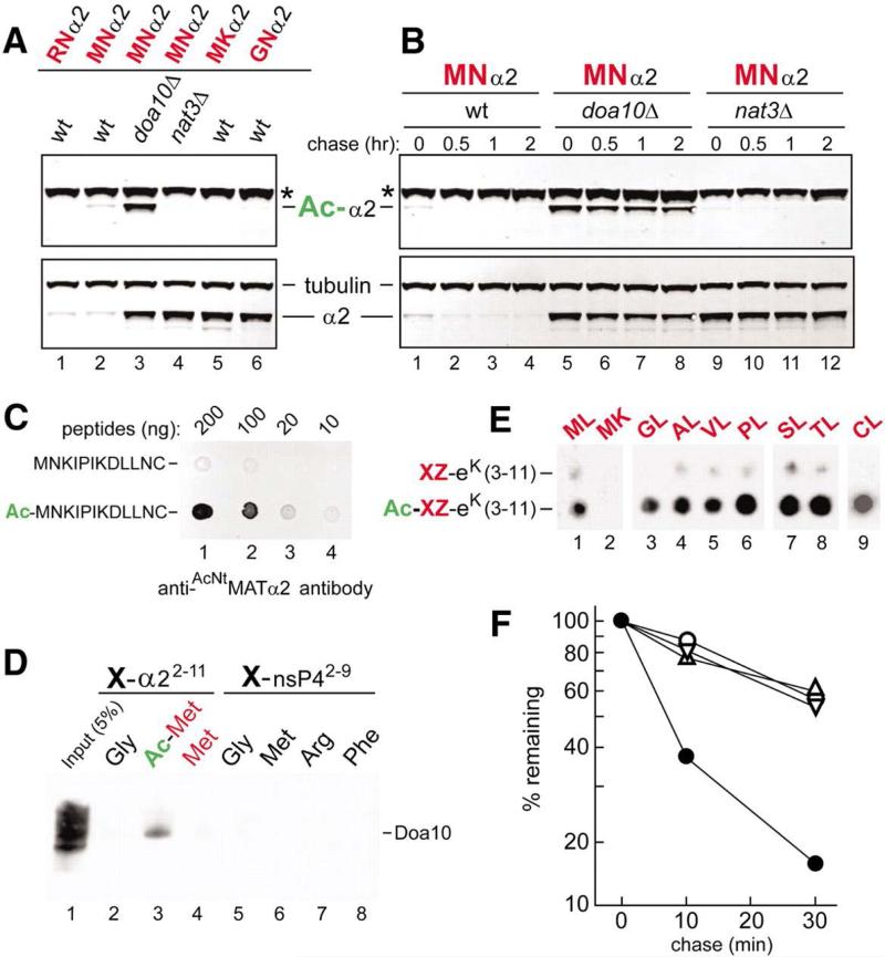
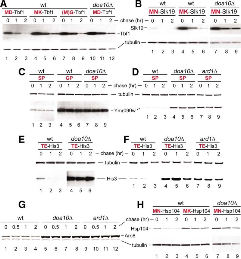
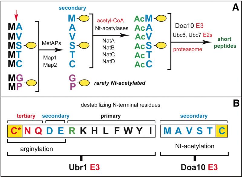
The retained N-terminal methionine (Met) residue of a nascent protein is often N-terminally acetylated (Nt-acetylated). Removal of N-terminal Met by Met-aminopeptidases frequently leads to Nt-acetylation of the resulting N-terminal alanine (Ala), valine (Val), serine (Ser), threonine (Thr), and cysteine (Cys) residues. Although a majority of eukaryotic proteins (for example, more than 80% of human proteins) are cotranslationally Nt-acetylated, the function of this extensively studied modification is largely unknown. Using the yeast Saccharomyces cerevisiae, we found that the Nt-acetylated Met residue could act as a degradation signal (degron), targeted by the Doa10 ubiquitin ligase. Moreover, Doa10 also recognized the Nt-acetylated Ala, Val, Ser, Thr, and Cys residues. Several examined proteins of diverse functions contained these N-terminal degrons, termed AcN-degrons, which are a prevalent class of degradation signals in cellular proteins.


Figures

Fig. 1
Fig. 1
Destabilizing N-terminal residues. (A) CHX chases, for 0, 1, and 2 hours in wild-typeS. cerevisiaeexpressing CK-eK-Ura3 (lanes 1 to 3) or CL-eK-Ura3 (lanes 4 to 6). Cell extracts were fractionated by SDS–polyacrylamide gel electrophoresis, followed by immunoblotting with anti-Ha and anti-tubulin, the latter a loading control. (B) As in (A) but chases for 0 and 2 hours with XZ-eK-Ura3 (X = Cys, Met, or Ser; Z = Trp, Val, or Leu) in wild-type versus ubr1Δ cells. (C) As in (A) but with MZ-eK-Ura3 (Z = Leu or Lys) in wild-type cells. (D) As in (A) but chases for 0, 0.5, and 1.5 hours with XL-eK-Ura3 (X = Gly, Val, Ala, or Thr) in wild-type cells. (E) As in (A) but with CL-eK-Ura3 in wild-type cells (lanes 1 to 3) versus doa10Δ cells (lanes 4 to 6). (F) As in (E) but chases for 0, 0.5, 1, and 2 hours with ML-eK-Ura3. (G) Lanes 1 and 2, short-lived ML-eK-Ura in the MG132-sensitive pdr5Δ S. cerevisiae, in the absence and presence of MG132, respectively. Lanes 3 and 4, same as lanes 1 and 2 but with long-lived MK-eK-Ura3.
Fig. 2
Fig. 2
Doa10 as an N-recognin. (A) Extracts from wild-type, doa10Δ, and nat3Δ S. cerevisiae that expressed XZ-α23-67-eK-Ura3 (XZα2) (X = Met, Arg, or Gly; Z = Asn or Lys) were immunoblotted with anti-AcNtMATα2 (which selectively recognized Nt-acetylated MNα2) or (separately) with anti-Ha, which recognized both Nt-acetylated and unacetylated MNα2, or with anti-tubulin. XZα2 (“α2”), Nt-acetylated XZα2 (“Ac-α2”), and tubulin are indicated. Asterisks denote a protein cross-reacting with anti-AcNtMATα2. (B) As in (A) but CHX chases for 0, 0.5, 1, and 2 hours with MNα2, in wild-type, doa10Δ, and nat3Δ cells. (C) Indicated amounts of the Ntacetylated Ac-MNKIPIKDLLNC peptide versus its unacetylated counterpart were spotted onto membrane and assayed for their binding to anti-AcNtMATα2. (D) X-peptide pulldown with peptides XNKIPIKDLLNC (X = Met, AcMet, or Gly) (lanes 2 to 4) or XIFSTDTGPGGC (X = Gly, Met, Arg, or Phe) (lanes 5 to 8) and extract of S. cerevisiae that expressed Doa10myc13. Lane 1, input extract (5%). (E) SPOT assay with purified, flag-tagged Doa10f and spot-arrayed synthetic peptides XZ-eK(3-11) (X = Gly, Ala, Val, Pro, Ser, Thr, or Cys; Z = Leu or Lys) and their Nt-acetylated XZ-eK(3-11)counterparts. XZ residues are indicated at the top of the membrane. (F) Quantitation, using a PhosphorImager, of 35S-pulse chases with MATα2f and its mutant derivatives (fig. S6, B and C). Solid circles, MNMATα2f; open circles,MKMATα2f; upright triangles, GNMATα2f (initially MGNMATα2f) in ubc4Δ cells; inverted triangles, MNMATα2f in ubc4Δ doa10Δ cells.
Fig. 3
Fig. 3
AcN-degrons in yeast proteins. (A) Lanes 1 to 3, CHX chase for 0, 1, and 2 hours in wild-type S. cerevisiaeexpressing Tbf1ha. Lanes 4 to 6, 7 to 9, and 10 to 12, analogous patterns but in doa10Δ cells withMKTbf1ha (Lys at position 2), GTbf1ha(initially MGTbf1ha), and wild-type Tbf1ha, respectively. (B) As in (A) but with wild-type Slk19ha and its mutant derivatives in wild-type versus doa10Δ cells. (C) As in (A) but with wild-type Ymr090wha and its mutant derivatives in wild-type versus doa10Δ cells. (D) As in (C) but with wild-type Ymr090wha in wild-type versus doa10Δ and ard1Δ cells. (E) As in (A) but with wild-type His3ha in wild-type versus doa10Δ cells. (F) As in (E), with wild-type His3ha in wild-type versus doa10Δ and ard1Δ cells. (G) As in (A) but CHX chases for 0, 0.5, 1, and 2 hours with wild-type Aro8hain wild-type versus doa10Δ and ard1Δ cells. (H) As in (A) but with wild-type Hsp104ha and its mutant derivatives in wild-type versus doa10Δ cells.
Fig. 4
Fig. 4
Nα-terminal acetylases, Met-aminopeptidases, and the Doa10 branch of the N-end–rule pathway. (A) The Doa10-mediated branch of the S. cerevisiaeN-end–rule pathway (see fig. S1C for the Ubr1-mediated branch of this pathway). The red arrow on the left indicates the MetAP-mediated removal of N-terminal Met. This Met is retained if a residue at position 2 is nonpermissive (too large) for MetAPs. If the retained N-terminal Met or N-terminal Ala, Val, Ser, Thr, and Cys are followed by acetylation-permissive residues, the above N-terminal residues are usually Ntacetylated (–7). The resulting N-degrons are termed AcN-degrons. The term “secondary” refers to the necessity of modification (Nt-acetylation) of a destabilizing N-terminal residue before a protein can be recognized by a cognate Ub ligase (fig. S1C). Proteins containing AcN-degrons are targeted for ubiquitylation (and proteasome-mediated degradation) by the Doa10 E3 Ub ligase. Although Gly or Pro can be made N-terminal by MetAPs, and although Doa10 can recognize Ntacetylated Gly and Pro (Fig. 2E), few proteins with N-terminal Gly or Pro are Nt-acetylated (– 7). (B) The Ubr1 and Doa10 branches of the N-end–rule pathway. Both branches target, through different mechanisms, the N-terminal Cys residue (yellow rectangles), with oxidized Cys marked by an asterisk.

Comment in

References

    1. Jörnvall H. J. Theor. Biol. 1975;55:1. - PubMed
    1. Mullen JR, et al. EMBO J. 1989;8:2067. - PMC - PubMed
    1. Park EC, Szostak JW. EMBO J. 1992;11:2087. - PMC - PubMed
    1. Gautschi M, et al. Mol. Cell. Biol. 2003;23:7403. - PMC - PubMed
    1. Polevoda B, Sherman F. J. Mol. Biol. 2003;325:595. - PubMed

Publication types

MeSH terms