. 2010 Jan 29;327(5965):580-3.
doi: 10.1126/science.1181928.

Platelets amplify inflammation in arthritis via collagen-dependent microparticle production

Affiliations

Platelets amplify inflammation in arthritis via collagen-dependent microparticle production

Eric Boilard et al. Science. .

Abstract

In addition to their pivotal role in thrombosis and wound repair, platelets participate in inflammatory responses. We investigated the role of platelets in the autoimmune disease rheumatoid arthritis. We identified platelet microparticles--submicrometer vesicles elaborated by activated platelets--in joint fluid from patients with rheumatoid arthritis and other forms of inflammatory arthritis, but not in joint fluid from patients with osteoarthritis. Platelet microparticles were proinflammatory, eliciting cytokine responses from synovial fibroblasts via interleukin-1. Consistent with these findings, depletion of platelets attenuated murine inflammatory arthritis. Using both pharmacologic and genetic approaches, we identified the collagen receptor glycoprotein VI as a key trigger for platelet microparticle generation in arthritis pathophysiology. Thus, these findings demonstrate a previously unappreciated role for platelets and their activation-induced microparticles in inflammatory joint diseases.


Figures

Fig. 1
Fig. 1
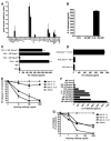
Platelet MPs are abundant in inflammatory SF. Cells in freshly isolated RA SF were stained with lineage markers: CD15 (neutrophils), CD3 (T cells), CD14 (monocytes and macrophages) and CD41 (platelets), or the appropriate isotype controls and analyzed by flow cytometry. (A) Forward- by side-scatter profiles of events in RA SF. Populations identified by further gating and lineage marker staining are labeled. (B) Representative histogram of CD41+ (black fill) platelet MPs resident in RA SF. Events were gated based on the forward-scatter parameters indicated in (A). (Gray fill, isotype control.) Data are representative of profiles from eight RA patients. (C) Flow cytometric quantification of CD41+ platelet MPs (<1 μm as determined by size calibration beads) in RA and OA SF after removal of leukocytes by centrifugation (n = 20 donors per group). (D) Flow cytometric quantification of platelet (CD41+) MPs in SF from juvenile idiopathic arthritis (JIA, n = 6), psoriatic arthritis (PA, n = 19), and gout (n = 14). (E) Flow cytometric quantification of MPs in RA SF derived from the indicated cell types (n = 19 donors).
Fig. 2
Fig. 2
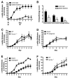
Platelets are involved in arthritis development. (A) Arthritis severity after K/BxN serum transfer in mice administered a platelet-depleting antibody (triangles) or isotype control (squares); n = 10 mice per group. Data are the mean ± SEM pooled from two independent experiments. P < 0.001. Arrows, parenteral administration of platelet-depleting antibody; arrowheads, K/BxN serum administration. (B) Histomorphometric quantification of arthritis severity in ankle joints of platelet-depleted and control mice at experiment termination; n = 10 mice per group. *P < 0.01. (C to F) Arthritis severity was measured after administration of K/BxN serum in mice (C) deficient in thromboxane synthase (Tbxas1−/−), treated daily with (D) the thromboxane A2 (TP) antagonist SQ 29,548 or (E) ADP:P2Y12 inhibitor clopidogrel, or in mice deficient in (F) GPIb (Gp1ba −/−). Data are the mean ± SEM, n = 10 mice per group. (C), (D), (F) P = not significant; (E) P < 0.001.
Fig. 3
Fig. 3
Platelets form MPs and participate in arthritis pathophysiology via stimulation of the collagen receptor GPVI. (A) Representative flow cytometry forward- and side-scatter plots of CD41+ mouse platelets incubated in the presence or absence of FLS. (B) Examination of candidate stimuli of murine platelet MP formation upon co-culture with FLS. Mouse platelets incubated in the presence of cyclooxygenase inhibitor salicylic acid (ASA), isolated from mice treated with ADP:P2Y12 inhibitor clopidogrel, or from the indicated gene-targeted mice were coincubated with mouse FLS. MP formation was quantified by flow cytometry. Data are the mean ± SEM, n = 3 independent experiments in duplicate. (C) Scanning electron micrograph of human platelets exhibiting MP budding when incubated in the presence of FLS. Arrows indicate the edge of the fibroblast-like synoviocyte. Upper and lower panels are 9800× and 69,270× magnifications, respectively. (D) Human platelets form MPs when incubated with FLS and when exposed to a GPVI-specific peptide ligand (CRP) but not in the presence of a related control peptide (GPP). (E) Arthritis severity after K/BxN serum transfer was quantified in GPVI-null (Gp6 −/−) (triangles) or wild-type control (squares) mice. Data are the mean ± SEM pooled from three independent experiments; n = 25 mice per group. P < 0.001. (F) Histomorphometric quantification of arthritis severity in ankle tissues from GPVI-null (Gp6 −/−) and WT mice at experimental termination. Data are the mean ± SEM; n = 25 mice per group. *P = 0.019, **P < 0.05, ***P < 0.01.
Fig. 4
Fig. 4
MPs activate FLS in an IL-1–dependent manner. (A) MPs generated by collagen stimulation of human platelets were coincubated with human FLS, and cytokine release was quantified by Proteome Profiler. Data are representative of three independent experiments. (B) MPs isolated from RA SF were coincubated with FLS, and supernatants were assayed for IL-8 release by ELISA (enzyme-linked immunosorbent assay). (C) Mouse platelet MPs generated by collagen stimulation of platelets from the indicated genotypes were coincubated with mouse FLS, and supernatants were assayed for KC release by ELISA. (D) Mouse MPs generated by collagen stimulation of WT platelets were coincubated with IL-1R1–null (Il1r1−/−) FLS, and supernatants were assayed for KC release by ELISA. Recombinant TNF (10 ng/ml) was added to FLS to induce KC release as a positive control. (E) Mouse platelet MPs were coincubated with FLS in the presence of IL-1–neutralizing antibodies, and supernatants were assayed for KC release by ELISA. (F) Potency of human MP stimulation of FLS. FLS were exposed to graded concentrations of IL-1β, TNF, or platelet MPs, and IL-8 release was quantified in culture supernatants by ELISA. (G) Human platelet MPs were coincubated with FLS in the presence of IL-1–neutralizing antibodies, and supernatants were assayed for IL-8 release by ELISA. Data for (B) to (G) are the mean ± SEM of three independent experiments.

Comment in

Similar articles

Cited by

References

    1. Gartner LP, Hiatt JL, Strum JM. BRS Cell Biology and Histology. 5. Lippincott Williams & Wilkins; Philadelphia, PA: 2006.
    1. George JN. Lancet. 2000;355:1531. - PubMed
    1. Davì G, Patrono C. N Engl J Med. 2007;357:2482. - PubMed
    1. Helmick CG, et al. Arthritis Rheum. 2008;58:15. - PubMed
    1. Lee DM, Weinblatt ME. Lancet. 2001;358:903. - PubMed

Publication types

MeSH terms