. 2010 Mar 19;106(5):971-80.
doi: 10.1161/CIRCRESAHA.109.210682. Epub 2010 Jan 28.

Relative roles of direct regeneration versus paracrine effects of human cardiosphere-derived cells transplanted into infarcted mice

Affiliations

Relative roles of direct regeneration versus paracrine effects of human cardiosphere-derived cells transplanted into infarcted mice

Isotta Chimenti et al. Circ Res. .

Abstract

Rationale: Multiple biological mechanisms contribute to the efficacy of cardiac cell therapy. Most prominent among these are direct heart muscle and blood vessel regeneration from transplanted cells, as opposed to paracrine enhancement of tissue preservation and/or recruitment of endogenous repair.

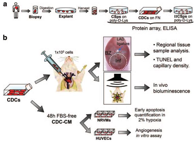
Objective: Human cardiac progenitor cells, cultured as cardiospheres (CSps) or as CSp-derived cells (CDCs), have been shown to be capable of direct cardiac regeneration in vivo. Here we characterized paracrine effects in CDC transplantation and investigated their relative importance versus direct differentiation of surviving transplanted cells.

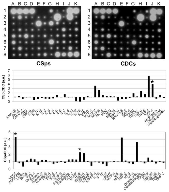
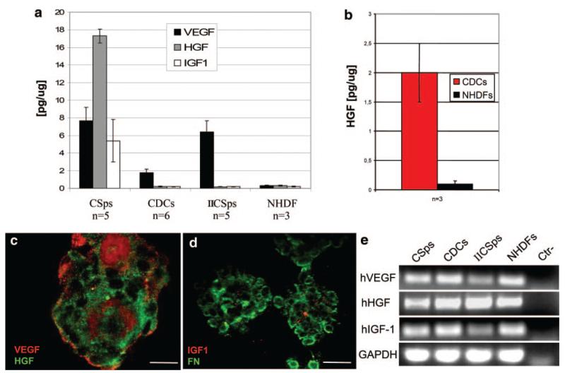
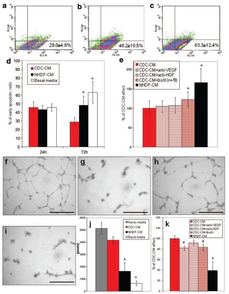
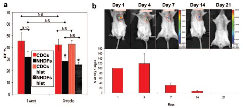
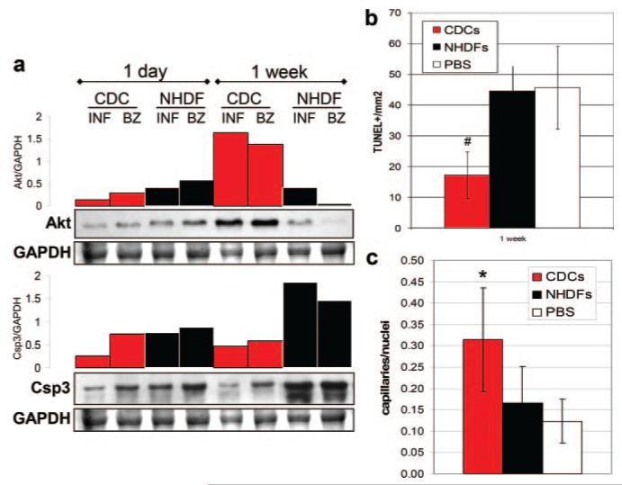
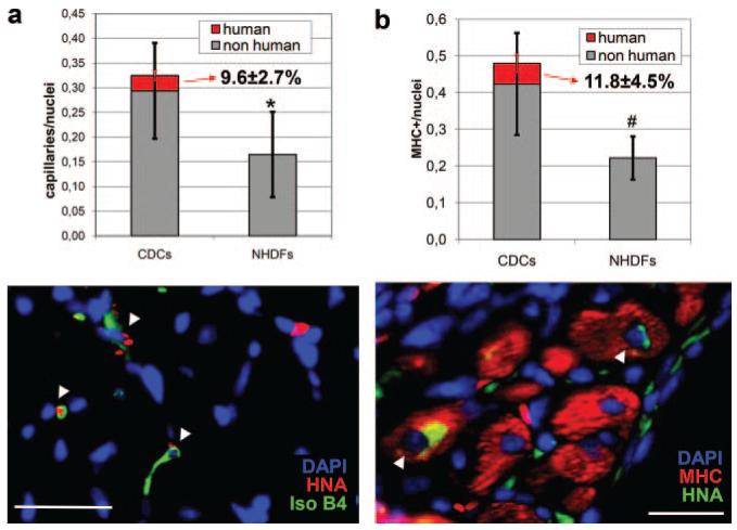
Methods and results: In vitro, many growth factors were found in media conditioned by human adult CSps and CDCs; CDC-conditioned media exerted antiapoptotic effects on neonatal rat ventricular myocytes, and proangiogenic effects on human umbilical vein endothelial cells. In vivo, human CDCs secreted vascular endothelial growth factor, hepatocyte growth factor, and insulin-like growth factor 1 when transplanted into the same SCID mouse model of acute myocardial infarction where they were previously shown to improve function and to produce tissue regeneration. Injection of CDCs in the peri-infarct zone increased the expression of Akt, decreased apoptotic rate and caspase 3 level, and increased capillary density, indicating overall higher tissue resilience. Based on the number of human-specific cells relative to overall increases in capillary density and myocardial viability, direct differentiation quantitatively accounted for 20% to 50% of the observed effects.

Conclusions: Together with their spontaneous commitment to cardiac and angiogenic differentiation, transplanted CDCs serve as "role models," recruiting endogenous regeneration and improving tissue resistance to ischemic stress. The contribution of the role model effect rivals or exceeds that of direct regeneration.


Figures

Figure 1
Figure 1. Experimental design
a, Culture protocol for CSps, CDCs, and IICSps isolation. b, Two sets of functional experiments were based on CDCs: in vivo injection of 1×10⁵ cells in a SCID mouse MI model for regional tissue samples analysis, histology, and luciferase detection; and in vitro assessment of CDC-CM effects on NRVM viability or HUVEC angiogenic abilities. CMs were collected after 48 hours from CDCs (or NHDFs as control) and used to plate NRVMs in 2% hypoxia or HUVECs on matrix-coated wells.
Figure 2
Figure 2. Cytokines and growth factors in vitro screening
Representative images of protein arrays on serum-free CM from CSps and CDCs, as well as corresponding densitograms of the CSp/CDC levels ratios for each spot. *Three selected factors of interest.
Figure 3
Figure 3. VEGF, HGF, and IGF1 secretion levels in vitro
CSps secrete VEGF, HGF, and IGF1 in vitro in low serum media, whereas in the same conditions, CDCs and IICSps only secrete VEGF (a). HGF release by CDCs is recovered in serum-free BM (b). Confocal images of CSps stained for VEGF and HGF (c) and IGF1 and fibronectin (FN) (d) allowed GF detection in their 3D structure. RT-PCR for GFs in CSps, CDCs, IICSps, and NHDFs is shown in e. Scale bars=100 μm.
Figure 4
Figure 4. Prosurvival and proangiogenic in vitro effects of CDC-CM
NRVMs were plated with CM in 2% hypoxia up to 72 hours. a through c, Representative density plots of early apoptotic NRVMs cultured for 72 hours in CDC-CM (n=11), NHDF-CM (n=11), or BM (n=7), respectively. Average percentages of 7AAD/Annexin V+ (FL1-H and FL3-H, respectively) are shown at the lower right of each graph. Data are summarized in graph d (n=5 per group at 24 hours). e, Preincubation of CDC-CM with VEGF and HGF neutralizing antibodies (n=19) significantly reduced viable NRVMs after 72 hours. f through i, Representative images of HUVECs 18 hours after plating on matrix-coated wells in endothelial cell media (f), serum-free BM (g), CMC-CM (h), and NHDF-CM (i). j, Quantification of total tube length in the different media. Preincubation of CDC-CM with VEGF neutralizing antibody significantly reduced the positive effect (k). *P<0.001, #P<0.05 vs CDC group. Scale bars=1 mm.
Figure 5
Figure 5. Human GFs are detectable in infarcted CDC-injected hearts
Human mRNAs for VEGF, HGF, and IGF1 are detectable in the infarcted tissue from murine hearts up to 3 weeks after LAD ligation and cell delivery, both in CDC- and NHDF-injected mice (a). For each time point, PCR products from 2 different animals are shown. Human VEGF, HGF, and IGF1 proteins are detectable with human specific antibodies in CDC-injected hearts at different time points, but not in NHDF-injected (b), although human GAPDH detection confirms engraftment in both groups. Inj indicates injected; INF, infarct; BZ, border zone; RV, right ventricle; SEP, septum.
Figure 6
Figure 6. Functional and engraftment evaluation in vivo
a, Comparison of LVEF in mice groups from the present study to 2 historical groups (hist) from a previous study. Data are plotted as averages±SEM. #P<0.05 vs the corresponding CDC group. NS: not statistically significant. b: Engraftment follow-up by in vivo bioluminescence on luciferase-labeled CDC injected mice. Signal intensity is normalized to that of day 1. Note that the representative images at 14 and 21 days have an expanded color scale bar.
Figure 7
Figure 7. Tissue viability and perfusion assessment at molecular and histological levels
a, WB for Akt and corresponding densitometric plots, normalized to GAPDH, showed that 1 week after surgery Akt expression was higher in infarct (INF) and BZ tissue from CDC-injected compared to NHDF-injected mice. Consistently, at the same time point, Csp3 protein levels were lower in CDC-injected hearts. b and c, Quantification of the TUNEL-positive cell density (b) and capillary density (c) in the BZ of infarcted mice 1 week after cell delivery. *P<0.001 and #P<0.05 vs control groups.
Figure 8
Figure 8. Human contribution to capillaries and MHC-positive cells in engraftment areas
a, Double staining for isolectin-B4 and HNA allowed assessment of CDC contribution to the overall capillary density in engrafted areas in the border zone. b, Double staining for MHC and HNA allowed quantification of direct muscle regeneration in the viable tissue areas of the infarct. *P<0.001 and #P<0.05 vs CDC group (in graph a refers to both overall counts and nonhuman only counts). Scale bars=25 μm. Arrowheads indicate double-positive cells.

Similar articles

Cited by

References

    1. Rosenzweig A. Cardiac cell therapy-mixed results from mixed cells. N Engl J Med. 2006;355:1274–1277. - PubMed
    1. Smith RR, Barile L, Messina E, Marban E. Stem cells in the heart: what’s the buzz all about? Part 1: preclinical considerations. Heart Rhythm. 2008;5:749–757. - PMC - PubMed
    1. Smith RR, Barile L, Messina E, Marban E. Stem cells in the heart: what’s the buzz all about? Part 2: arrhythmic risks and clinical studies. Heart Rhythm. 2008;5:880–887. - PMC - PubMed
    1. Dimmeler S, Burchfield J, Zeiher AM. Cell-based therapy of myocardial infarction. Arterioscler Thromb Vasc Biol. 2008;28:208–216. - PubMed
    1. Guan K, Hasenfuss G. Do stem cells in the heart truly differentiate into cardiomyocytes? J Mol Cell Cardiol. 2007;43:377–387. - PubMed

ONLINE SUPPLEMENTAL REFERENCES

    1. Smith RR, Barile L, Cho HC, Leppo MK, Hare JM, Messina E, Giacomello A, Abraham MR, Marban E. Regenerative potential of cardiosphere-derived cells expanded from percutaneous endomyocardial biopsy specimens. Circulation. 2007;115:896–908. - PubMed
    1. Iravanian S, Nabutovsky Y, Kong CR, Saha S, Bursac N, Tung L. Functional reentry in cultured monolayers of neonatal rat cardiac cells. Am J Physiol Heart Circ Physiol. 2003;285:H449–456. - PubMed
    1. Kizana E, Ginn SL, Allen DG, Ross DL, Alexander IE. Fibroblasts can be genetically modified to produce excitable cells capable of electrical coupling. Circulation. 2005;111:394–398. - PubMed
    1. Zhang YW, Su Y, Lanning N, Gustafson M, Shinomiya N, Zhao P, Cao B, Tsarfaty G, Wang LM, Hay R, Vande Woude GF. Enhanced growth of human met-expressing xenografts in a new strain of immunocompromised mice transgenic for human hepatocyte growth factor/scatter factor. Oncogene. 2005;24:101–106. - PubMed
    1. Zeng F, Chen MJ, Baldwin DA, Gong ZJ, Yan JB, Qian H, Wang J, Jiang X, Ren ZR, Sun D, Huang SZ. Multiorgan engraftment and differentiation of human cord blood CD34+ Lin-cells in goats assessed by gene expression profiling. Proc atl Acad Sci U S A. 2006;103:7801–7806. - PMC - PubMed

Publication types

MeSH terms