. 2010 Mar 5;106(4):686-94.
doi: 10.1161/CIRCRESAHA.109.205732. Epub 2010 Jan 28.

Decreased levels of embryonic retinoic acid synthesis accelerate recovery from arterial growth delay in a mouse model of DiGeorge syndrome

Affiliations

Decreased levels of embryonic retinoic acid synthesis accelerate recovery from arterial growth delay in a mouse model of DiGeorge syndrome

Lucile Ryckebüsch et al. Circ Res. .

Abstract

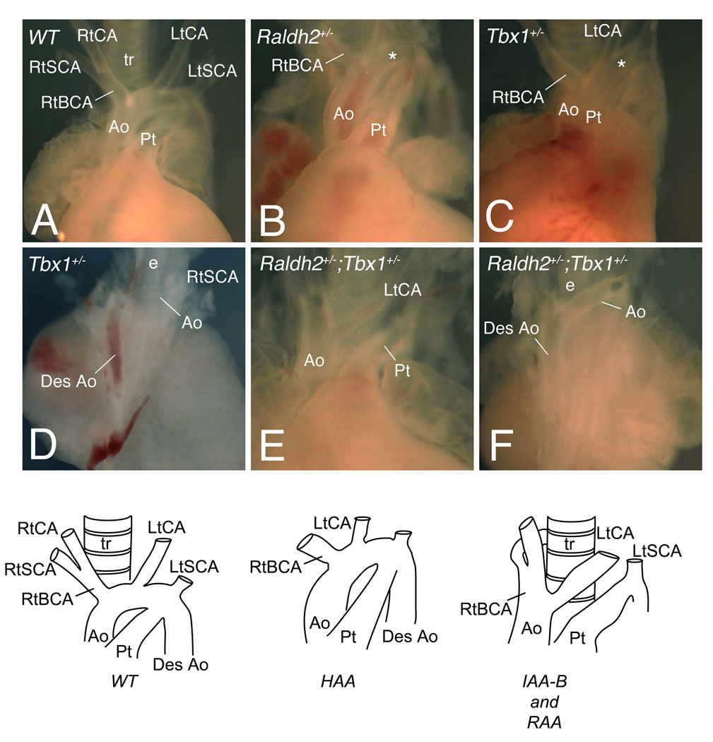
Rationale: Loss of Tbx1 and decrease of retinoic acid (RA) synthesis result in DiGeorge/velocardiofacial syndrome (DGS/VCFS)-like phenotypes in mouse models, including defects in septation of the outflow tract of the heart and anomalies of pharyngeal arch-derived structures including arteries of the head and neck, laryngeal-tracheal cartilage, and thymus/parathyroid. Wild-type levels of T-box transcription factor (Tbx)1 and RA signaling are required for normal pharyngeal arch artery development. Recent studies have shown that reduction of RA or loss of Tbx1 alters the contribution of second heart field (SHF) progenitor cells to the elongating heart tube.

Objective: Here we tested whether Tbx1 and the RA signaling pathway interact during the deployment of the SHF and formation of the mature aortic arch.

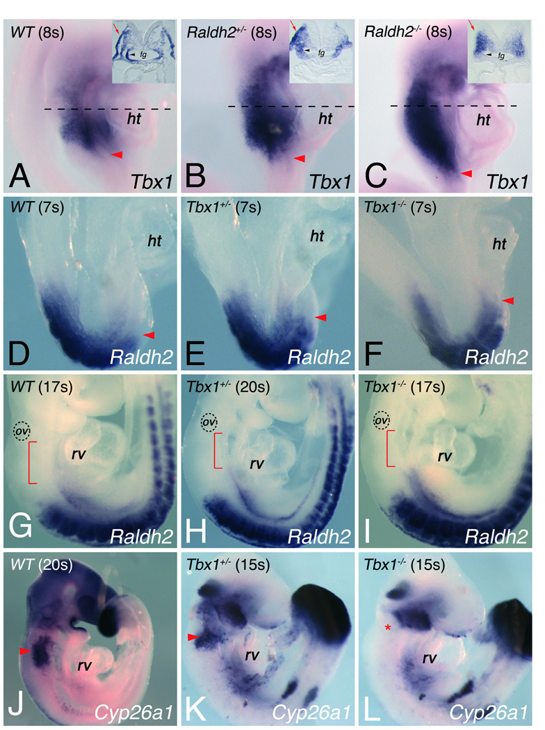
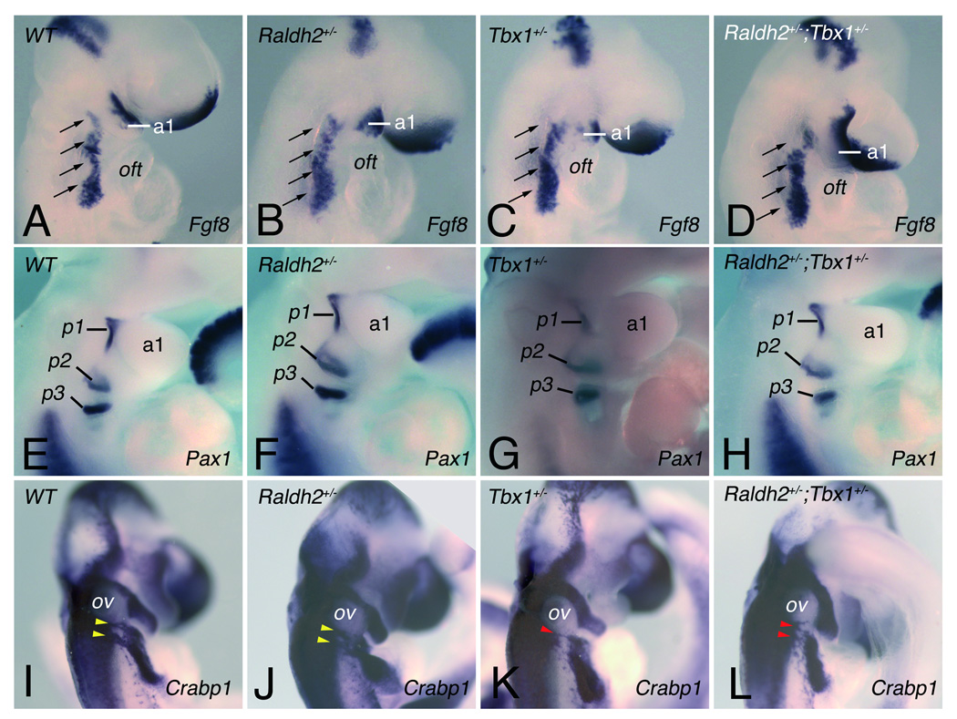
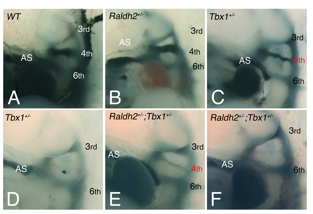
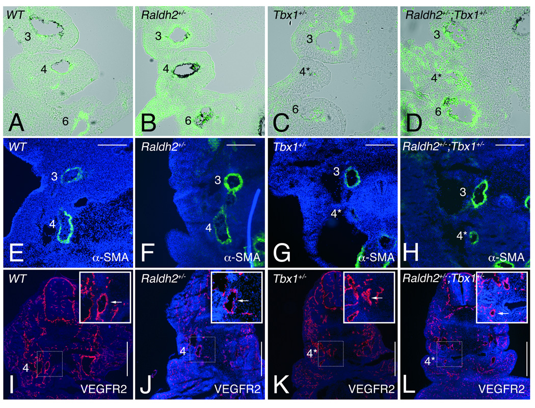
Methods and results: Molecular markers of the SHF, neural crest and smooth muscle cells, were analyzed in Raldh2;Tbx1 compound heterozygous mutants. Our results revealed that the SHF and outflow tract develop normally in Raldh2(+/-);Tbx1(+/-) embryos. However, we found that decreased levels of RA accelerate the recovery from arterial growth delay observed in Tbx1(+/-) mutant embryos. This compensation coincides with the differentiation of smooth muscle cells in the 4th pharyngeal arch arteries, and is associated with severity of neural crest cell migration defects observed in these mutants.

Conclusions: Our data suggest that differences in levels of embryonic RA may contribute to the variability in great artery anomalies observed in DGS/VCFS patients.


Figures

Figure 1
Figure 1
Alteration of Tbx1 and retinoic acid signaling in Raldh2 and Tbx1 mutant embryos. In situ hybridization on whole mount embryos shows (A-C) Tbx1, (D-I) Raldh2 and (J-L) Cyp26a1 (I,J) in different mutant backgrounds. (A-C) Red arrowheads indicate the most posterior limit of Tbx1 expression in (A) wild-type (WT), (B) Raldh2+/− and (C) Raldh2−/− embryos at the 8-somite stage (8s), dotted lines indicating planes of sections. Transverse sections of embryos displayed in insets in A-C, indicate that RA signaling is required for Tbx1 transcript accumulation in pharyngeal ectoderm (red arrows) and endoderm (black arrowheads). Tbx1 expression is expanded caudally in absence of Raldh2 (compare C with A). (D-I) Raldh2 expression in (D,G) WT, (E,H) Tbx1+/− and (F,I) Tbx1−/− embryos at the 7-somite stage (7s) and at embryonic day (E) 9.5. Arrowheads in D-F show the anterior limit of Raldh2 expression. Red brackets in G-I indicate the interval between the most anterior domain of Raldh2 expression and the otic vesicle (ov). (J-L) In situ hybridization showing Cyp26a1 expression in (J) WT, (K) Tbx1+/− and (L) Tbx1−/− embryos at 15s stage, revealing the absence of Cyp26a1 expression in ectoderm caudal to pharyngeal brachial two (asterisk) in absence of Tbx1−/− embryos (compare L with K). fg, foregut; ht, heart tube; rv, right ventricle.
Figure 2
Figure 2
Pharyngeal and neural crest development in single and compound heterozygous Tbx1 and Raldh2 mutant embryos. (A-D) Fgf8 expression in the pharyngeal region at E9.5 detected by in situ hybridization in (A) WT, (B) Raldh2+/−, (C) Tbx1+/− and (D) Raldh2+/−;Tbx1+/− embryos. Analysis of Fgf8 (A-D) transcripts displays no difference between single and compound heterozygous embryos. Arrows indicate the pharyngeal region where Fgf8 is expressed. (E-F) Endodermal pouch formation was assessed by expression of Pax1 detected at E10.5 (~30–34 somites). Pharyngeal pouches 1–3 were bilaterally present in all embryos (see Online Figure II for the fourth pharyngeal pouch). (I-L) Migratory cardiac neural crest cells (NCC) were detected by ISH for Crabp1 in WT, in single and compound heterozygous embryos at E10.5. Yellow and red arrowheads indicate normal and abnormal post-otic streams of migrating NCCs, respectively. a, arch; oft, outflow tract; ov, otic vesicle; p, pouch.
Figure 3
Figure 3
Pharyngeal arch artery (PAA) abnormalities visualized by intracardiac injection of India ink at E10.5. (A-F) Left lateral views of injected embryos displaying normal formation of the left 3rd, 4th and 6th PAAs in (A) WT and (B) Raldh2+/− embryos, whereas a defect of one or both 4th PAAs is observed in (C,D) Tbx1+/− and (E,F) compound heterozygous embryos. Defects include hypoplasia (red numbers) in (C) Tbx1+/− and (E) Raldh2+/−;Tbx1+/− embryos, or absence of the 4th PAAs in (D) Tbx1+/− and (F) Raldh2+/−;Tbx1+/− embryos. See Table 1 for detail. Arabic numerals indicate PAAs. AS, aortic sac.
Figure 4
Figure 4
Vascular smooth muscle differentiation in single and compound heterozygous Tbx1 and Raldh2 mutant embryos. (A-H) Vascular smooth muscle (VSM) differentiation was followed by anti-αSMA on front sections through the pharyngeal region of embryos at (A-D) E10.5, and (E-H) E11.5. (A-D) Abnormally small 4th PAAs (asterisk in C) in Tbx1+/− (n=6) embryos at E10.5 were devoid of VSM staining, while weak staining was detected (asterisk in D) in Raldh2+/−,Tbx1+/− (n=8) embryos at the same stage. At E11.5 4th PAAs of (G) Tbx1+/− embryos had a thin and incomplete layer of VSM in the vessel wall (n=5), while normal VSM differentiation was observed in the 4th PAAs of (H) Raldh2+/−;Tbx1+/− embryos (n=6). (I-L) Immunofluorescent analysis of VEGFR2 of the 4th PAAs in front sections of (I) WT, (J) Raldh2+/−, (K) Tbx1+/− and (L) Raldh2+/−;Tbx1+/− embryos at E11.5. Insets show high magnification views of the 4th PAA (arrow) as indicated by the boxed areas in I-L. VEGFR2 was not affected in hypoplatic 4th PAAs (asterisk) in (K) Tbx1+/− and (L) Raldh2+/−;Tbx1+/− embryos. See Table 1 and Table 2 for details. l, left; r, right. Scale bars: 200µm (E through H); 500µm (I through L)
Figure 5
Figure 5
Aortic arch abnormalities in single and compound heterozygous Tbx1 and Raldh2 mutants at fetal stages. (A,B,C,E) Ventral or (D,F) dorsal views of dissected fetal hearts showing the great arteries of WT and mutant embryos. (A) WT embryo showing a left aortic arch and normal origin of the right subclavian and left carotid arteries at E14.5. Examples of (B) Raldh2+/− and (C) Tbx1+/− embryos with a high aortic arch (asterisk). The dorsal views (D,F) show a retro-esophageal connection between the ascending and descending aorta (desAo) in Tbx1+/− and compound Raldh2+/−;Tbx1+/− embryos respectively. The frontal view (E) reveals lack of segment of the arch, a type B interruption (IAA-B) in Raldh2+/−;Tbx1+/− mutant. Schemas represent a WT, high aortic arch (HAA) and interruption (IAA-B) with retro-esophageal connection respectively. See Table 3 for detail. Ao, aorta; BCA, brachiocephalic artery; CA, carotid artery; e, esophagus; Lt, left; Pt, pulmonary trunk; Rt, right; SCA, subclavian artery; t, trachea.

Comment in

References

    1. Poelmann RE, Gittenberger-de Groot AC, Hierck BP. The development of the heart and microcirculation: role of shear stress. Med Biol Eng Comput. 2008;46:479–484. - PMC - PubMed
    1. Yashiro K, Shiratori H, Hamada H. Haemodynamics determined by a genetic programme govern asymmetric development of the aortic arch. Nature. 2007;450:285–288. - PubMed
    1. Lindsay EA. Chromosomal microdeletions: dissecting del22q11 syndrome. Nat Rev Genet. 2001;2:858–868. - PubMed
    1. Baldini A. Dissecting contiguous gene defects: TBX1. Curr Opin Genet Dev. 2005;15:279–284. - PubMed
    1. Hu T, Yamagishi H, Maeda J, McAnally J, Yamagishi C, Srivastava D. Tbx1 regulates fibroblast growth factors in the anterior heart field through a reinforcing autoregulatory loop involving forkhead transcription factors. Development. 2004;131:5491–5502. - PubMed

Publication types

MeSH terms