

Gonadal mRNA expression levels of TGFbeta superfamily signaling factors correspond with post-hatching morphological development in American alligators
- PMID: 20110644
- PMCID: PMC2855286
- DOI: 10.1159/000277934
Gonadal mRNA expression levels of TGFbeta superfamily signaling factors correspond with post-hatching morphological development in American alligators
Abstract
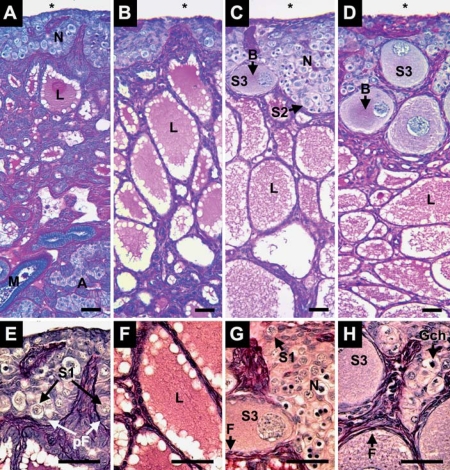
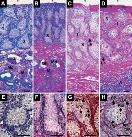
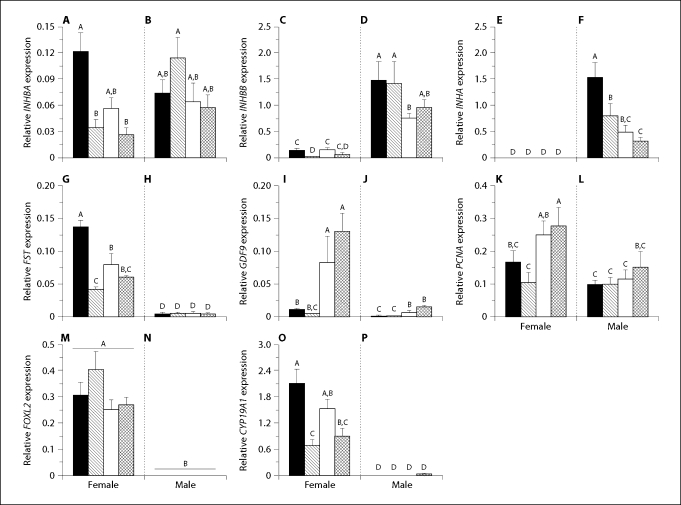
Paracrine factor signaling regulates many aspects of vertebrate gonadal development. We investigated key ovarian and testicular morphological markers of the American alligator (Alligator mississippiensis) during the first 5 months post-hatching and correlated gonadal development with mRNA expression levels of a suite of regulatory factors. In both sexes, we observed significant morphology changes, including ovarian follicle assembly and meiotic progression of testicular germ cells. Concomitant with these changes were sexually dimorphic and ontogenetically variable mRNA expressions. In ovaries, FOXL2, aromatase, and follistatin mRNA expression was greater than in testes at all ages. At one week after hatching, we observed ovarian medullary remodeling in association with elevated activin/inhibin beta A subunit, follistatin, and aromatase mRNA expressions. Three and 5 months following hatching and concomitant with follicle assembly, ovaries showed increased mRNA expression levels of GDF9 and the mitotic factor PCNA. In testes, the activin/inhibin alpha and beta B subunit transcript levels were greater than in ovaries at all ages. Elevated testicular expression of GDF9 mRNA levels at 5 months after hatching aligned with increased spermatogenic activity. We propose that the mRNA expression levels and concomitant morphological changes observed here affect the establishment of alligator reproductive health and later fertility.
(c) 2010 S. Karger AG, Basel.
Figures
Similar articles
-
Altered sex hormone concentrations and gonadal mRNA expression levels of activin signaling factors in hatchling alligators from a contaminated Florida lake.J Exp Zool A Ecol Genet Physiol. 2010 Apr 1;313(4):218-30. doi: 10.1002/jez.595. J Exp Zool A Ecol Genet Physiol. 2010. PMID: 20166196 Free PMC article.
-
Gonadotropin-induced changes in oviducal mRNA expression levels of sex steroid hormone receptors and activin-related signaling factors in the alligator.Gen Comp Endocrinol. 2012 Jan 15;175(2):251-8. doi: 10.1016/j.ygcen.2011.11.018. Epub 2011 Dec 1. Gen Comp Endocrinol. 2012. PMID: 22154572 Free PMC article.
-
Posthatching development of Alligator mississippiensis ovary and testis.J Morphol. 2010 May;271(5):580-95. doi: 10.1002/jmor.10818. J Morphol. 2010. PMID: 20013789 Free PMC article.
-
Altered gonadal expression of TGF-β superfamily signaling factors in environmental contaminant-exposed juvenile alligators.J Steroid Biochem Mol Biol. 2011 Oct;127(1-2):58-63. doi: 10.1016/j.jsbmb.2011.01.004. Epub 2011 Jan 18. J Steroid Biochem Mol Biol. 2011. PMID: 21251980
-
Activins in reproductive biology and beyond.Hum Reprod Update. 2016 Apr;22(3):342-57. doi: 10.1093/humupd/dmv058. Epub 2016 Feb 15. Hum Reprod Update. 2016. PMID: 26884470 Review.
Cited by
-
Altered sex hormone concentrations and gonadal mRNA expression levels of activin signaling factors in hatchling alligators from a contaminated Florida lake.J Exp Zool A Ecol Genet Physiol. 2010 Apr 1;313(4):218-30. doi: 10.1002/jez.595. J Exp Zool A Ecol Genet Physiol. 2010. PMID: 20166196 Free PMC article.
-
Gene-environment interactions: the potential role of contaminants in somatic growth and the development of the reproductive system of the American alligator.Mol Cell Endocrinol. 2012 May 6;354(1-2):111-20. doi: 10.1016/j.mce.2011.10.020. Epub 2011 Oct 28. Mol Cell Endocrinol. 2012. PMID: 22061623 Free PMC article.
-
Gonadotropin-induced changes in oviducal mRNA expression levels of sex steroid hormone receptors and activin-related signaling factors in the alligator.Gen Comp Endocrinol. 2012 Jan 15;175(2):251-8. doi: 10.1016/j.ygcen.2011.11.018. Epub 2011 Dec 1. Gen Comp Endocrinol. 2012. PMID: 22154572 Free PMC article.
-
Identification and evaluation of antioxidant and reference genes for quantitative real-time PCR in blood of Caiman latirostris.Heliyon. 2021 Feb 13;7(2):e06253. doi: 10.1016/j.heliyon.2021.e06253. eCollection 2021 Feb. Heliyon. 2021. PMID: 33659756 Free PMC article.
-
Estrogen receptor 1 (ESR1; ERα), not ESR2 (ERβ), modulates estrogen-induced sex reversal in the American alligator, a species with temperature-dependent sex determination.Endocrinology. 2015 May;156(5):1887-99. doi: 10.1210/en.2014-1852. Epub 2015 Feb 25. Endocrinology. 2015. PMID: 25714813 Free PMC article.
References
-
- Barakat B, O'Connor AE, Gold E, de Kretser DM, Loveland KL. Inhibin, activin, follistatin and FSH serum levels and testicular production are highly modulated during the first spermatogenic wave in mice. Reproduction. 2008;136:345–359. - PubMed
-
- Belaid B, Richard-Mercier N, Pieau C, Dorizzi M. Sex reversal and aromatase in the European pond turtle: treatment with letrozole after the thermosensitive period for sex determination. J Exp Zool. 2001;290:490–497. - PubMed
-
- Billiar RB, Zachos NC, Burch MG, Albrecht ED, Pepe GJ. Up-regulation of alpha-inhibin expression in the fetal ovary of estrogen-suppressed baboons is associated with impaired fetal ovarian folliculogenesis. Biol Reprod. 2003;68:1989–1996. - PubMed
-
- Bristol-Gould SK, Kreeger PK, Selkirk CG, Kilen SM, Cook RW, et al. Postnatal regulation of germ cells by activin: The establishment of the initial follicle pool. Dev Biol. 2006;298:132–148. - PubMed
Publication types
MeSH terms
Substances
Grants and funding
LinkOut - more resources
Full Text Sources
Miscellaneous
