. 2009 Dec 30;15(1):114-27.
doi: 10.3390/molecules15010114.

JNK contributes to Hif-1alpha regulation in hypoxic neurons

Affiliations

JNK contributes to Hif-1alpha regulation in hypoxic neurons

Xanthi Antoniou et al. Molecules. .

Abstract

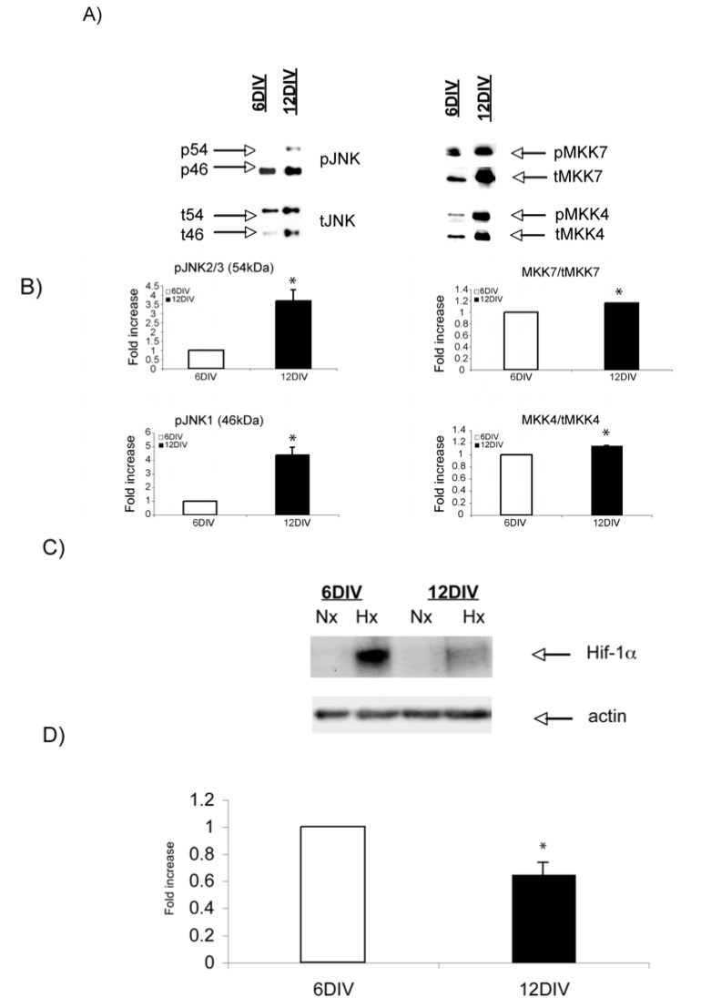
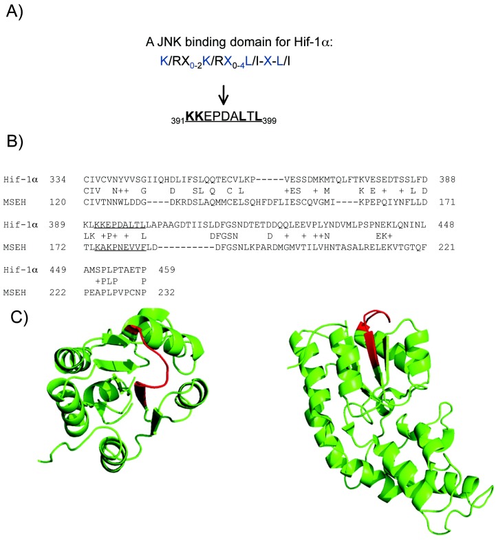
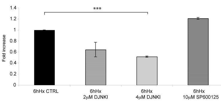
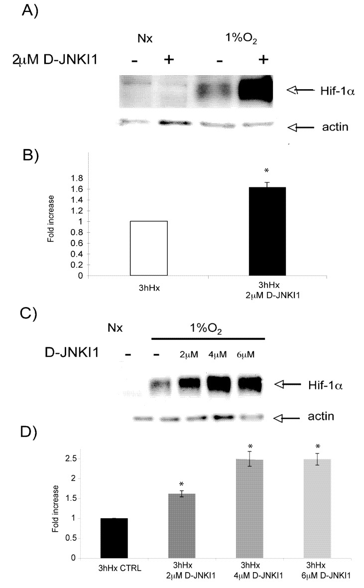
Hypoxia is an established factor of neurodegeneration. Nowadays, attention is directed at understanding how alterations in the expression of stress-related signaling proteins contribute to age dependent neuronal vulnerability to injury. The purpose of this study was to investigate how Hif-1alpha, a major neuroprotective factor, and JNK signaling, a key pathway in neurodegeneration, relate to hypoxic injury in young (6DIV) and adult (12DIV) neurons. We could show that in young neurons as compared to mature ones, the protective factor Hif-1alpha is more induced while the stress protein phospho-JNK displays lower basal levels. Indeed, changes in the expression levels of these proteins correlated with increased vulnerability of adult neurons to hypoxic injury. Furthermore, we describe for the first time that treatment with the D-JNKI1, a JNK-inhibiting peptide, rescues adult hypoxic neurons from death and contributes to Hif-1alpha upregulation, probably via a direct interaction with the Hif-1alpha protein.


Figures

Figure 1
Figure 1
Characterisation of the model: A) 12DIV neurons are more susceptible to hypoxic injury. Hypoxia leads to a significant increase in neuronal death by 6h in adult neurons (p < 0.001). Young neurons are resistant to hypoxic injury for prolonged periods. B) Representative Western blot images of the NMDARs protein levels: NR1, NR2A, NR2B. C) Quantification of Western blots shows significant increase of all three receptor subtypes with increasing age (p < 0.001). D) Application of the NMDA blocker MK-801 (5 μM) rescues adult neurons from hypoxic neuronal death (p < 0.001). Data are presented as fold increase in comparison to normoxic untreated controls. Quantification is from three independent experiments.
Figure 2
Figure 2
Basal levels of JNK signaling proteins increase with increasing age, while Hif-1α hypoxic induction decreases. (A) Representative Western blot images of phospho-JNK (pJNK), and its regulators pMKK7 and pMMK4 in 6DIV and 12DIV neurons. Basal levels of pJNK, pMKK7 and pMKK4 are higher in adult 12DIV neurons. (B) Quantification confirms significant up regulation of the JNK signaling components with increasing age (p < 0.05). (C) Representative Western blot images of Hif-1αhypoxic induction in 6DIV and 12DIV neurons. (D) Quantification of Western blots shows that Hif-1α hypoxic induction is 0.4 fold ± 0.086 lower in 12DIV adult neurons (p < 0.05). Data are presented as fold increase in comparison to normoxic controls. Quantification is from three independent experiments.
Figure 3
Figure 3
pJNK and Hif-1α are regulated by hypoxia at the protein level. A) Western blot analysis shows that pJNK is transiently induced by hypoxia, but falls below normoxic levels by 2h. B) Quantification confirms a biphasic regulation of pJNK2/3 (Significant downregulation by 30 min (p < 0.05)). pJNK1 was significantly increased by 30 min (p < 0.05). C) Western blot analysis reveals that Hif-1α expression is induced by hypoxia in a time-dependent manner. D) Quantification of Western blots shows that Hif-1α is significantly increased by 1h and reaches a peak by 3h (p < 0.001). Data are presented as fold increase in comparison to normoxic controls. Quantification is from three independent experiments.
Figure 4
Figure 4
Computational evidence indicates Hif-1α as a potential target for JNK. A) Hif-1α contains a potential JNK binding domain (JBD) within the N-terminal VHL (Von Hippel-Lindau) recognition site. The JBD motif, as well the corresponding JBD sequence within Hif-1α are indicated. B) The local sequence alignment between MSEH and Hif-1α, the JBD motif and its corresponding sequence in MSEH are underlined. C) Cartoon representation of the 3D structure of MSEH (pdb code 1CQZ), the sequence corresponding to the JBD motif are coloured in red; at the left, view from the top and at the right, view from the side.
Figure 5
Figure 5
D-JNKI1 treatment rescues neurons from hypoxic induced death. D-JNKI1 treatment of adult neurons prior to hypoxic exposure led to a significant decrease in neuronal death, as assessed by LDH assay release. 2 μM D-JNKI1 led to a significant decrease of neuronal death, while 4 μM further decreased neuronal death (p < 0.001). Application of SP600125 (10 μM) prior to hypoxia did not revert neuronal death. Data are presented as fold increase in comparison to hypoxic controls. Quantification is from three independent experiments.
Figure 6
Figure 6
D-JNKI1 increases hypoxia-induced Hif-1αexpression. A) Representative Western blot of Hif-1α after hypoxic exposure shows D-JNKI1 (2 μΜ) increases hypoxia-induced Hif-1α expression. B) Quantification analysis confirmed that hypoxia-induced Hif-1α expression is significantly increased following D-JNKI1 treatment (p < 0.05). C) Representative Western Blot shows that hypoxia-induced Hif-1αexpression increases by D-JNKI1 in a dose dependent manner. D) Quantification confirmed significant upregulation of Hif-1α expression with increasing D-JNKI1 concentrations in comparison to hypoxic untreated controls (p < 0.05). Data are presented as fold increase in comparison to hypoxic untreated controls. Quantification is from three independent experiments.

Similar articles

Cited by

References

    1. Munns S.E., Meloni B.P., Knuckey N.W., Arthur P.G. Primary cortical neuronal cultures reduce cellular energy utilization during anoxic energy deprivation. J. Neurochem. 2003;87:764–772. doi: 10.1046/j.1471-4159.2003.02049.x. - DOI - PubMed
    1. Mattson M.P., Magnus T. Ageing and neuronal vulnerability. Nat. Rev. Neurosci. 2006;7:278–294. doi: 10.1038/nrn1886. - DOI - PMC - PubMed
    1. Loerch P.M., Lu T., Dakin K.A., Vann J.M., Isaacs A., Geula C., Wang J., Pan Y., Gabuzda D.H., Li C., Prolla T.A., Yankner B.A. Evolution of the aging brain transcriptome and synaptic regulation. PLoS One. 2008;3:e3329. - PMC - PubMed
    1. Lu T., Pan Y., Kao S.Y., Li C., Kohane I., Chan J., Yankner B.A. Gene regulation and DNA damage in the ageing human brain. Nature. 2004;429:883–891. - PubMed
    1. Ogunshola O.O., Antoniou X. Contribution of hypoxia to Alzheimer's disease: Is HIF-1alpha a mediator of neurodegeneration? Cell. Mol. Life Sci. 2009;66:22. - PMC - PubMed

Publication types

Substances