

Aberrant methylation inactivates transforming growth factor Beta receptor I in head and neck squamous cell carcinoma
- PMID: 20111589
- PMCID: PMC2809419
- DOI: 10.1155/2009/848695
Aberrant methylation inactivates transforming growth factor Beta receptor I in head and neck squamous cell carcinoma
Abstract
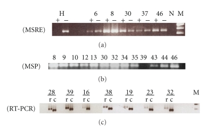
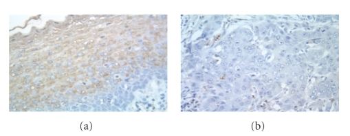
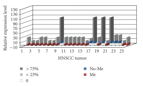
Background. Alterations in TGF-beta signaling are common in head and neck cancer (HNSCC). Mutations in TGF-beta type II receptor (TbetaR-II) occur frequently in HNSCC while TGF-beta type I receptor (TbetaR-I) mutations are rare, suggesting that other molecular alterations in the TGF-beta pathway are likely. To identify abnormalities in TbetaR-I expression we analyzed 50 HNSCCs and correlated the results with clinical-pathologic features. Methods. Hypermethylation of TbetaR-I was evaluated via methylation-specific PCR (MSP) and restriction enzyme-mediated PCR (MSRE). Mutations in exons 1 and 7, mRNA and protein expression were analyzed by direct sequencing, semiquantitative RT-PCR and immunohistochemistry, respectively. Results. TbetaR-I expression was lost in 83% HNSCCs and was linked to DNA hypermethylation of the CpG-rich promoter region in 62% of the tumors. The variants 9A/6A and Int7G24A were found in two patients. Conclusions. This study shows that suppression of TbetaR-I expression in HNSCC is associated with DNA hypermethylation.
Figures
Similar articles
-
Defective transforming growth factor beta signaling pathway in head and neck squamous cell carcinoma as evidenced by the lack of expression of activated Smad2.Clin Cancer Res. 2001 Jun;7(6):1618-26. Clin Cancer Res. 2001. PMID: 11410498
-
Expression of transforming growth factor beta type II receptors in head and neck squamous cell carcinoma.Clin Cancer Res. 1999 Jun;5(6):1243-8. Clin Cancer Res. 1999. PMID: 10389906
-
Frequent alterations of Smad signaling in human head and neck squamous cell carcinomas: a tissue microarray analysis.Oncol Res. 2003;14(2):61-73. doi: 10.3727/000000003108748612. Oncol Res. 2003. PMID: 14649540
-
Transforming growth factor beta receptors in verrucous and squamous cell carcinoma.Arch Otolaryngol Head Neck Surg. 1999 Aug;125(8):849-54. doi: 10.1001/archotol.125.8.849. Arch Otolaryngol Head Neck Surg. 1999. PMID: 10448730
-
TGF-beta control of cell proliferation.J Cell Biochem. 2005 Oct 15;96(3):447-62. doi: 10.1002/jcb.20558. J Cell Biochem. 2005. PMID: 16088940 Review.
Cited by
-
Epigenetic Alteration by DNA Promoter Hypermethylation of Genes Related to Transforming Growth Factor-β (TGF-β) Signaling in Cancer.Cancers (Basel). 2011 Mar 3;3(1):982-93. doi: 10.3390/cancers3010982. Cancers (Basel). 2011. PMID: 24212650 Free PMC article.
-
Advances and Challenges in Targeting TGF-β Isoforms for Therapeutic Intervention of Cancer: A Mechanism-Based Perspective.Pharmaceuticals (Basel). 2024 Apr 20;17(4):533. doi: 10.3390/ph17040533. Pharmaceuticals (Basel). 2024. PMID: 38675493 Free PMC article. Review.
-
Role of DNA methylation in head and neck cancer.Clin Epigenetics. 2011 Aug;2(2):123-50. doi: 10.1007/s13148-011-0045-3. Epub 2011 Jul 9. Clin Epigenetics. 2011. PMID: 22704334 Free PMC article.
-
Transforming Growth Factor Β as a Marker of Hepatocellular Carcinoma in Patients with Chronic Hepatitis C Virus Infection.Rep Biochem Mol Biol. 2023 Jan;11(4):702-709. doi: 10.52547/rbmb.11.4.702. Rep Biochem Mol Biol. 2023. PMID: 37131893 Free PMC article.
-
Array CGH analysis of the rare laryngeal basaloid squamous cell carcinoma: a case report.Int J Clin Exp Pathol. 2012;5(8):834-9. Epub 2012 Oct 1. Int J Clin Exp Pathol. 2012. PMID: 23071866 Free PMC article.
References
-
- Myers EN, Suen JY, Myers J, Hanna E. Cancer of the Head and Neck. 4th edition. Philadelphia, Pa, USA: W. B. Saunders; 2003.
-
- American Cancer Society. Cancer Facts and Figures 2007. Atlanta, Ga, USA: American Cancer Society; 2007.
-
- Patel SG, Shah JP. TNM staging of cancers of the head and neck: striving for uniformity among diversity. CA: A Cancer Journal for Clinicians. 2005;55(4):242–258. - PubMed
-
- Báez A, Almodóvar JI, Cantor A, et al. High frequency of HPV16-associated head and neck squamous cell carcinoma in the Puerto Rican population. Head and Neck. 2004;26(9):778–784. - PubMed
-
- Kreimer AR, Clifford GM, Boyle P, Franceschi S. Human papillomavirus types in head and neck squamous cell carcinomas worldwide: a systemic review. Cancer Epidemiology Biomarkers and Prevention. 2005;14(2):467–475. - PubMed
Grants and funding
LinkOut - more resources
Full Text Sources
Research Materials
