

Middle ear glandular neoplasms: adenoma, carcinoma or adenoma with neuroendocrine differentiation: a case series
- PMID: 20111612
- PMCID: PMC2812983
- DOI: 10.1186/1757-1626-0002-0000006508
Middle ear glandular neoplasms: adenoma, carcinoma or adenoma with neuroendocrine differentiation: a case series
Abstract
Introduction: Middle ear glandular neoplasms are infrequent causes of a middle ear mass. They can have exocrine and/or neuroendocrine differentiation. It is currently thought that these tumors are indistinguishable each from another. Herein, we present a new case of a middle ear glandular neoplasm. Our objective is to review all cases described in the literature in order to identify the clinical features, the gross pathology, the histopathology, the immunohistochemistry, to discuss the differential diagnosis, the treatment, the rate of recurrence, the follow-up, the incidence of metastasis, the prognosis and to present a new classification of middle ear glandular neoplasm.
Methods: We performed a MEDLINE database search for MEA-related articles published between 1950 and March 2008. The information from the reports was analyzed.
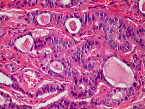
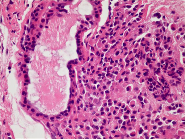
Results: Ninety-four patients with a middle ear adenoma are included in this report. We uncovered 75 patients with a carcinoid tumor and 19 patients with a middle ear adenoma diagnosis; the most common presenting symptom was a conductive hearing loss. Middle ear adenomas are lesions that are typically white, gray or reddish brown. They are grossly vascular and well circumscribed, but not encapsulated, and can entrap and destroy the ossicles. Histologically, the cuboidal to low columnar cells are arranged in a solid, trabecular or glandular architecture. The tumor's cells are immunohistochemically positive for a variety of keratin antibodies and most of them are also positive for neuroendocrine markers. Surgical excision is the treatment of choice. Local recurrence following complete excision is quite uncommon and metastases are rare.
Conclusions: Our study and the review of the literature showed adenomas and carcinoid tumors of the middle ear to be essentially indistinguishable benign tumors with metastatic potential. Based on the presence or absence of immunohistochemical markers and metastasis, we have classified these lesions into three types. Complete surgical treatment is recommended with an indefinite follow-up for possible recurrence.
Figures


Similar articles
-
Adenoma versus carcinoid tumor of the middle ear: a study of 48 cases and review of the literature.Mod Pathol. 2002 May;15(5):543-55. doi: 10.1038/modpathol.3880561. Mod Pathol. 2002. PMID: 12011260
-
Carcinoid tumor of the middle ear: clinical features, recurrences, and metastases.Laryngoscope. 2005 Sep;115(9):1660-6. doi: 10.1097/01.mlg.0000175069.13685.37. Laryngoscope. 2005. PMID: 16148713 Review.
-
'Neuroendocrine' middle ear adenomas: consistent expression of the transcription factor ISL1 further supports their neuroendocrine derivation.Histopathology. 2015 Jan;66(2):182-91. doi: 10.1111/his.12447. Epub 2014 Nov 10. Histopathology. 2015. PMID: 24766278
-
[Middle ear adenoma: clinical and pathologic analysis].Zhonghua Bing Li Xue Za Zhi. 2015 Dec;44(12):900-4. Zhonghua Bing Li Xue Za Zhi. 2015. PMID: 26888509 Chinese.
-
Middle ear adenoma/carcinoid tumour: a case report and review of the literature.Rev Laryngol Otol Rhinol (Bord). 2009;130(3):199-202. Rev Laryngol Otol Rhinol (Bord). 2009. PMID: 20345079 Review.
Cited by
-
A rare finding of pulmonary nodules in a middle ear neuroendocrine tumor: a case report and review of the literature.J Surg Case Rep. 2024 Oct 21;2024(10):rjae662. doi: 10.1093/jscr/rjae662. eCollection 2024 Oct. J Surg Case Rep. 2024. PMID: 39435307 Free PMC article.
-
Middle Ear Neuroendocrine Adenoma: A Case Report and Literature Review.Case Rep Otolaryngol. 2020 Dec 21;2020:8863188. doi: 10.1155/2020/8863188. eCollection 2020. Case Rep Otolaryngol. 2020. PMID: 33425416 Free PMC article.
-
Neuroendocrine adenoma of the middle ear-An intriguing entity.Clin Case Rep. 2021 Jan 25;9(3):1358-1361. doi: 10.1002/ccr3.3767. eCollection 2021 Mar. Clin Case Rep. 2021. PMID: 33768844 Free PMC article.
-
Neuroendocrine adenoma of the middle ear presented as paragaglioma: a case report.Hippokratia. 2017 Oct-Dec;21(4):201-203. Hippokratia. 2017. PMID: 30944513 Free PMC article.
-
Neuroendocrine neoplasms of the middle ear: report of 2 cases and review of the literature.Int J Clin Exp Pathol. 2019 Sep 1;12(9):3453-3458. eCollection 2019. Int J Clin Exp Pathol. 2019. PMID: 31934190 Free PMC article.
References
-
- Murphy GF, Pilch BZ, Dickersin GR, Goodman ML, Nadol JB Jr. Carcinoid tumor of the middle ear. Am J Clin Pathol. 1980;73:816–23. - PubMed
-
- Wassef M, Kanavaros P, Nemeth J, Adjamagbo H, Ba Huy PT. Amphicrine adenoma of the middle ear. Histological, immunohistochemical and ultrastructural study of a case. Ann Pathol. 1993;13:170–5. - PubMed
LinkOut - more resources
Full Text Sources
