

The role of bloom index of gelatin on the interaction with retinal pigment epithelial cells
- PMID: 20111679
- PMCID: PMC2812822
- DOI: 10.3390/ijms10083442
The role of bloom index of gelatin on the interaction with retinal pigment epithelial cells
Abstract
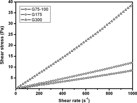
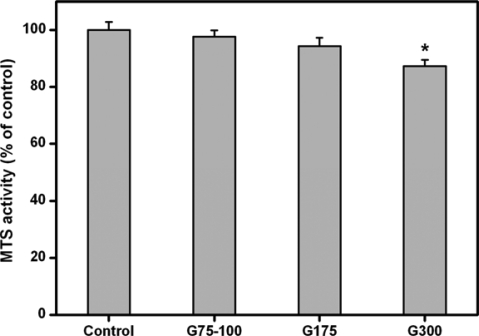
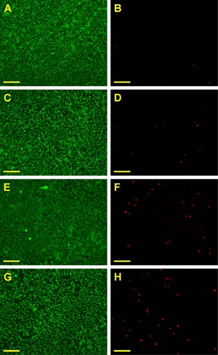
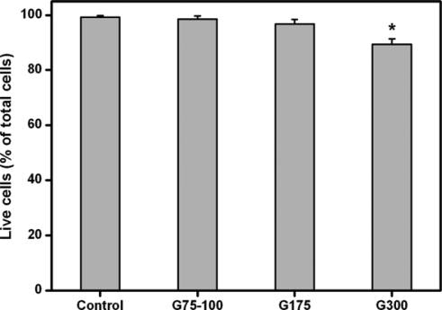
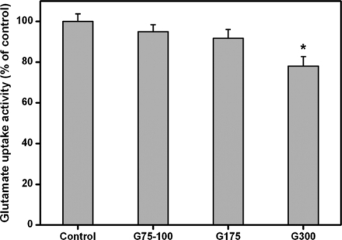
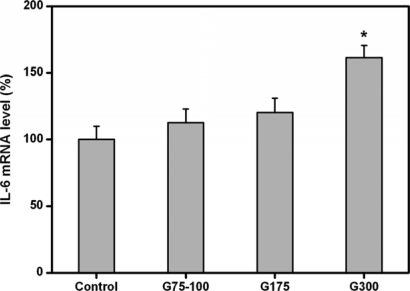
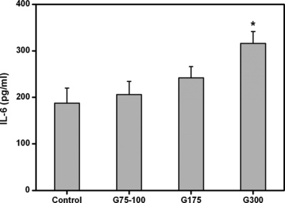
Biocompatible materials are of considerable interest in the development of cell/drug delivery carriers for therapeutic applications. This paper investigates the effects of the Bloom index of gelatin on its interaction with retinal pigment epithelial (RPE) cells. Following two days of culture of ARPE-19 cells with gelatin samples G75-100, G175, and G300, the in vitro biocompatibility was determined by cell proliferation and viability assays, and glutamate uptake measurements, as well as cytokine expression analyses. The mitochondrial dehydrogenase activity in the G300 groups was significantly lower than that of G75-100 and G175 groups. The Live/Dead assays also showed that the gelatin samples G300 induced mild cytotoxicity. In comparison with the treatment of gelatins with low Bloom index, the exposure to high Bloom strength gelatins markedly reduced the glutamate uptake capacity of ARPE-19 cells. One possible explanation for these observations is that the presence of gelatin samples G300 with high viscosity in the medium may affect the nutrient availability to cultured cells. The analyses of pro-inflammatory cytokine IL-6 expression at both mRNA and protein levels showed that the gelatins with low Bloom index caused less cellular inflammatory reaction and had more acceptable biocompatibility than their high Bloom strength counterparts. These findings suggest that the Bloom index gives influence on cellular responses to gelatin materials.
Keywords: Bloom index; gelatin; in vitro biocompatibility; retinal pigment epithelial cells.
Figures
Similar articles
-
Low Bloom strength gelatin as a carrier for potential use in retinal sheet encapsulation and transplantation.Biomacromolecules. 2009 Feb 9;10(2):310-9. doi: 10.1021/bm801039n. Biomacromolecules. 2009. PMID: 19063667
-
Effect of charge and molecular weight on the functionality of gelatin carriers for corneal endothelial cell therapy.Biomacromolecules. 2006 Jun;7(6):1836-44. doi: 10.1021/bm0601575. Biomacromolecules. 2006. PMID: 16768405
-
Influence of Cross-Linker Concentration on the Functionality of Carbodiimide Cross-Linked Gelatin Membranes for Retinal Sheet Carriers.J Biomater Sci Polym Ed. 2011;22(1-3):277-95. doi: 10.1163/092050609X12603600753204. J Biomater Sci Polym Ed. 2011. PMID: 20557713
-
Biocompatibility of chemically cross-linked gelatin hydrogels for ophthalmic use.J Mater Sci Mater Med. 2010 Jun;21(6):1899-911. doi: 10.1007/s10856-010-4035-3. Epub 2010 Mar 18. J Mater Sci Mater Med. 2010. PMID: 20238149
-
Mechanical properties and biocompatibility of in situ enzymatically cross-linked gelatin hydrogels.Int J Artif Organs. 2017 May 9;40(4):159-168. doi: 10.5301/ijao.5000553. Epub 2017 Mar 18. Int J Artif Organs. 2017. PMID: 28315501
Cited by
-
Nanoscale modification of porous gelatin scaffolds with chondroitin sulfate for corneal stromal tissue engineering.Int J Nanomedicine. 2012;7:1101-14. doi: 10.2147/IJN.S28753. Epub 2012 Feb 23. Int J Nanomedicine. 2012. PMID: 22403490 Free PMC article.
-
Modified Fish Gelatin as an Alternative to Mammalian Gelatin in Modern Food Technologies.Polymers (Basel). 2020 Dec 19;12(12):3051. doi: 10.3390/polym12123051. Polymers (Basel). 2020. PMID: 33352683 Free PMC article. Review.
-
Gelatin as It Is: History and Modernity.Int J Mol Sci. 2023 Feb 10;24(4):3583. doi: 10.3390/ijms24043583. Int J Mol Sci. 2023. PMID: 36834993 Free PMC article. Review.
-
Biotechnological Preparation of Gelatines from Chicken Feet.Polymers (Basel). 2019 Jun 18;11(6):1060. doi: 10.3390/polym11061060. Polymers (Basel). 2019. PMID: 31216750 Free PMC article.
-
Biological Evaluation of Thermosensitive Hydrogels of Chitosan/Hydrolyzed Collagen/β-GP in an In Vitro Model of Induced Cardiac Ischemia.Polymers (Basel). 2024 Aug 2;16(15):2206. doi: 10.3390/polym16152206. Polymers (Basel). 2024. PMID: 39125232 Free PMC article.
References
-
- Djagny KB, Wang Z, Xu S. Gelatin: A valuable protein for food and pharmaceutical industries: review. Crit. Rev. Food Sci. Nutr. 2001;41:481–492. - PubMed
-
- Bigi A, Cojazzi G, Panzavolta S, Rubini K, Roveri N. Mechanical and thermal properties of gelatin films at different degrees of glutaraldehyde crosslinking. Biomaterials. 2001;22:763–768. - PubMed
-
- Tabata Y, Ikada Y. Protein release from gelatin matrices. Adv. Drug Deliv. Rev. 1998;31:287–301. - PubMed
-
- Bussemer T, Dashevsky A, Bodmeier R. A pulsatile drug delivery system based on rupturable coated hard gelatin capsules. J. Control. Release. 2003;93:331–339. - PubMed
-
- Fan H, Zhang C, Li J, Bi L, Qin L, Wu H, Hu Y. Gelatin microspheres containing TGF-β3 enhance the chondrogenesis of mesenchymal stem cells in modified pellet culture. Biomacromolecules. 2008;9:927–934. - PubMed
Publication types
MeSH terms
Substances
LinkOut - more resources
Full Text Sources
Other Literature Sources
