

Reduced prefrontal gyrification in obsessive-compulsive disorder
- PMID: 20112027
- PMCID: PMC2940041
- DOI: 10.1007/s00406-009-0096-z
Reduced prefrontal gyrification in obsessive-compulsive disorder
Abstract
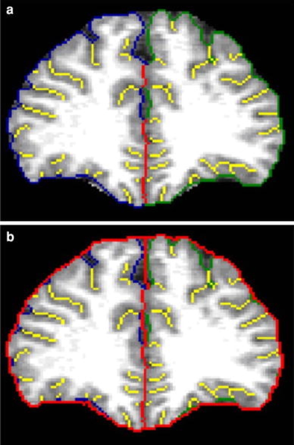
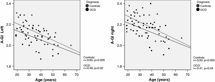
Structural magnetic resonance imaging (MRI) studies reveal evidence for brain abnormalities in obsessive-compulsive disorder (OCD), for instance, reduction of gray matter volume in the prefrontal cortex. Disturbances of gyrification in the prefrontal cortex have been described several times in schizophrenia pointing to a neurodevelopmental etiology, while gyrification has not been studied so far in OCD patients. In 26 OCD patients and 38 healthy control subjects MR-imaging was performed. Prefrontal cortical folding (gyrification) was measured bilaterally by an automated version of the automated-gyrification index (A-GI), a ratio reflecting the extent of folding, from the slice containing the inner genu of the corpus callosum up to the frontal pole. Analysis of covariance (ANCOVA, independent factor diagnosis, covariates age, duration of education) demonstrated that compared with control subjects, patients with OCD displayed a significantly reduced A-GI in the left hemisphere (p = 0.021) and a trend for a decreased A-GI in the right hemisphere (p = 0.076). Significant correlations between prefrontal lobe volume and A-GI were only observed in controls, but not in OCD patients. In conclusion, prefrontal hypogyrification in OCD patients may be a structural correlate of the impairment in executive function of this patient group and may point to a neurodevelopmental origin of this disease.
Figures
Similar articles
-
Corpus callosal signal intensity in treatment-naive pediatric obsessive compulsive disorders.Prog Neuropsychopharmacol Biol Psychiatry. 1999 May;23(4):601-12. doi: 10.1016/s0278-5846(99)00019-6. Prog Neuropsychopharmacol Biol Psychiatry. 1999. PMID: 10390719
-
Association of cortical gyrification, white matter microstructure, and phenotypic profile in medication-naïve obsessive-compulsive disorder.Psychol Med. 2024 Jun;54(8):1573-1579. doi: 10.1017/S0033291723003422. Epub 2023 Nov 23. Psychol Med. 2024. PMID: 37994452
-
Hypogyrification in obsessive-compulsive disorder.Psychol Med. 2017 Apr;47(6):1053-1061. doi: 10.1017/S0033291716003202. Epub 2016 Dec 12. Psychol Med. 2017. PMID: 27938423
-
Modality-specific overlaps in brain structure and function in obsessive-compulsive disorder: Multimodal meta-analysis of case-control MRI studies.Neurosci Biobehav Rev. 2020 May;112:83-94. doi: 10.1016/j.neubiorev.2020.01.033. Epub 2020 Jan 30. Neurosci Biobehav Rev. 2020. PMID: 32006553 Review.
-
Reduced focal fiber collinearity in the cingulum bundle in adults with obsessive-compulsive disorder.Neuropsychopharmacology. 2019 Jun;44(7):1182-1188. doi: 10.1038/s41386-019-0353-4. Epub 2019 Feb 25. Neuropsychopharmacology. 2019. PMID: 30802896 Free PMC article. Review.
Cited by
-
Functional significance of atypical cortical organization in spina bifida myelomeningocele: relations of cortical thickness and gyrification with IQ and fine motor dexterity.Cereb Cortex. 2013 Oct;23(10):2357-69. doi: 10.1093/cercor/bhs226. Epub 2012 Aug 8. Cereb Cortex. 2013. PMID: 22875857 Free PMC article.
-
Study on symptom dimensions and clinical characteristics in patients with obsessive-compulsive disorder.Rev Assoc Med Bras (1992). 2024 Mar 15;70(1):e20230676. doi: 10.1590/1806-9282.20230676. eCollection 2024. Rev Assoc Med Bras (1992). 2024. PMID: 38511751 Free PMC article.
-
Evidence for alterations of cortical folding in anorexia nervosa.Eur Arch Psychiatry Clin Neurosci. 2017 Feb;267(1):41-49. doi: 10.1007/s00406-015-0666-1. Epub 2015 Dec 17. Eur Arch Psychiatry Clin Neurosci. 2017. PMID: 26678081
-
Association between hippocampus volume and symptom profiles in obsessive-compulsive disorder.Neuroimage Clin. 2017 Nov 8;17:474-480. doi: 10.1016/j.nicl.2017.11.006. eCollection 2018. Neuroimage Clin. 2017. PMID: 29159060 Free PMC article.
-
A numerical simulation of neural fields on curved geometries.J Comput Neurosci. 2018 Oct;45(2):133-145. doi: 10.1007/s10827-018-0697-5. Epub 2018 Oct 11. J Comput Neurosci. 2018. PMID: 30306384 Free PMC article.
References
Publication types
MeSH terms
LinkOut - more resources
Full Text Sources
Medical
