. 2010 May;67(9):1505-18.
doi: 10.1007/s00018-010-0264-3. Epub 2010 Jan 29.

The human alpha(2)-plasmin inhibitor: functional characterization of the unique plasmin(ogen)-binding region

Affiliations

The human alpha(2)-plasmin inhibitor: functional characterization of the unique plasmin(ogen)-binding region

Simon S Gerber et al. Cell Mol Life Sci. 2010 May.

Abstract

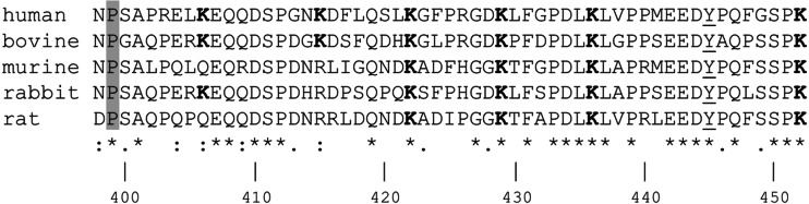
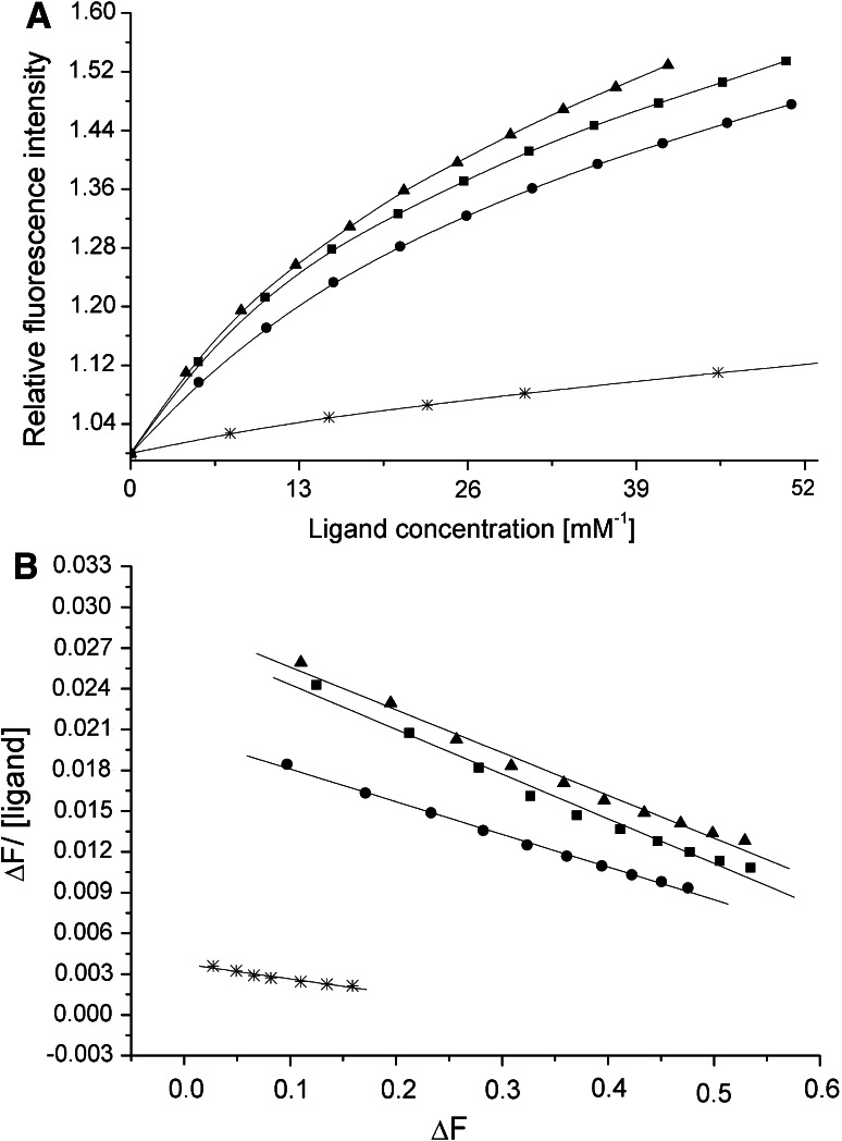
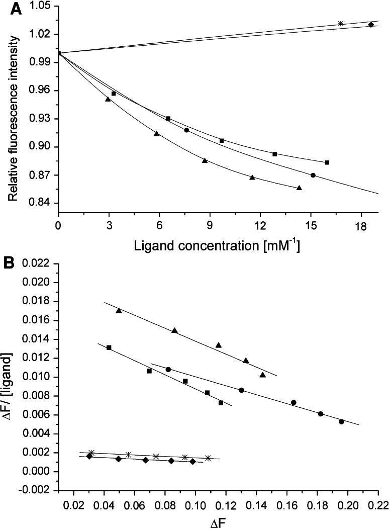
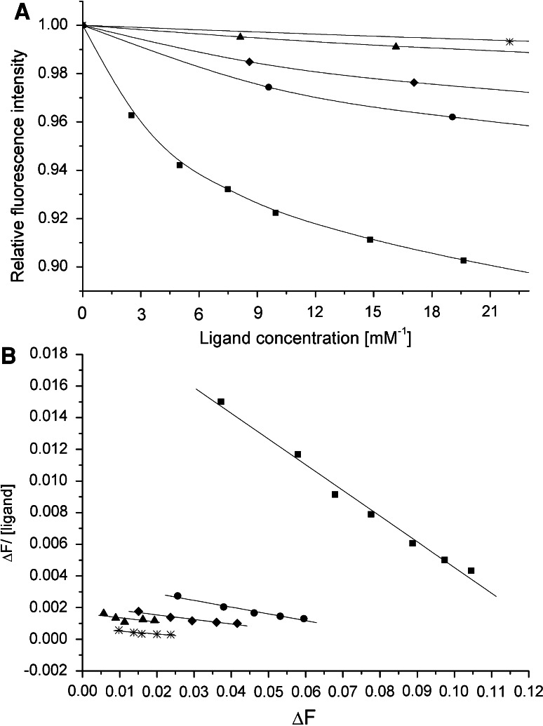
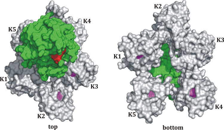
The human alpha(2)-plasmin inhibitor (A2PI) possesses unique N- and C-terminal extensions that significantly influence its biological activities. The C-terminal segment, A2PIC (Asn(398)-Lys(452)), contains six lysines thought to be involved in the binding to lysine-binding sites in the kringle domains of human plasminogen, of which four (Lys(422), Lys(429), Lys(436), Lys(452)) are completely and two (Lys(406), Lys(415)) are partially conserved. Multiple Lys to Ala mutants of A2PIC were expressed in Escherichia coli and used in intrinsic fluorescence titrations with kringle domains K1, K4, K4 + 5, and K1 + 2 + 3 of human plasminogen. We were able to identify the C-terminal Lys(452) as the main binding partner in recombinant A2PIC (rA2PIC) constructs with isolated kringles. We could show a cooperative, zipper-like enhancement of the interaction between C-terminal Lys(452) and internal Lys(436) of rA2PIC and isolated K1 + 2 + 3, whereas the other internal lysine residues contribute only to a minor extent to the binding process. Sulfated Tyr(445) in the unique C-terminal segment revealed no influence on the binding affinity to kringle domains.


Figures

Scheme 1
Scheme 1
Multiple sequence alignment of human, bovine, murine, rabbit, and rat A2PIC. The numbering is according to the Asn form of human A2PI. The Pro399Ala mutation is shaded in gray, sulfated Tyr445 is underlined, Lys residues are in bold type and the symbols asterisk, colon, dot, and blank space stand for identity, high, average, and poor similarity, respectively
Fig. 1
Fig. 1
a Averaged fluorescence titration of K4 with wild-type rA2PIC (filled squares), single Lys to Ala mutants rA2PIC(Lys436Ala) (filled triangles), and rA2PIC(Lys452Ser) (star), and quintuple mutant rA2PIC(Lys406Ala/Lys415Ala/Lys422Ala/Lys429Ala/Lys436Ala) (filled circles). The fluorescence intensity is normalized to 1 and the x-axis is enlarged. b Corresponding Scatchard plots and calculated regression curves
Fig. 2
Fig. 2
a Averaged fluorescence titration of rK1 with wild-type rA2PIC (filled squares), single Lys to Ala mutants rA2PIC(Lys436Ala) (filled triangles), and rA2PIC(Lys452Ser) (star), double rA2PIC(Lys429Ala/Lys452Ala) (filled diamonds), and quintuple rA2PIC(Lys406Ala/Lys415Ala/Lys422Ala/Lys429Ala/Lys436Ala) (filled circles) mutants. The fluorescence intensity is normalized to 1 and the x-axis is enlarged. b Corresponding Scatchard plots and calculated regression curves
Fig. 3
Fig. 3
a Averaged fluorescence titration of K1–3 with wild-type rA2PIC (filled squares), single Lys to Ala mutants rA2PIC(Lys436Ala) (filled triangles) and rA2PIC(Lys452Ser) (star), double rA2PIC(Lys429Ala/Lys452Ala) (filled diamonds), and quintuple rA2PIC(Lys406Ala/Lys415Ala/Lys422Ala/Lys429Ala/Lys436Ala) (filled circles) mutants. The fluorescence intensity is normalized to 1 and the x-axis is enlarged. b Corresponding Scatchard plots and calculated regression curves
Fig. 4
Fig. 4
Model structure of the whole Pgn molecule. The model is based on the amino acid sequence of Pgn, which was used as a template for the automated structure modeling using SWISS-MODEL [–59]. Connected and overlapping 3D structures of Pgn fragments (K1–K2; Leu81-Thr245, parts of K3–K4; Thr291-Ser441, parts of K4–K5; Leu402-Ala543, K1–K2–K3; Leu81-Cys333, and K5; Asp461-Pro544) were selected and thereafter assembled and attached to the 3D structure of the catalytic domain of Plm (1BML) using the visualization and rendering software PyMOL. Each kringle domain (K) is shown in gray, the central Trp of each LBS is presented in pink, the protease domain is colored in green, and the active site residues are marked in red

Similar articles

Cited by

References

    1. Lijnen HR, Wiman B, Collen D. Partial primary structure of human alpha 2-antiplasmin-homology with other plasma protease inhibitors. Thromb Haemost. 1982;48:311–314. - PubMed
    1. Collen D. Identification and some properties of a new fast-reacting plasmin inhibitor in human plasma. Eur J Biochem. 1976;69:209–216. doi: 10.1111/j.1432-1033.1976.tb10875.x. - DOI - PubMed
    1. Moroi M, Aoki N. Isolation and characterization of α2-plasmin inhibitor from human plasma. J Biol Chem. 1976;251:5956–5965. - PubMed
    1. Müllertz S, Clemmensen I. The primary inhibitor of plasmin in human plasma. Biochem J. 1976;159:545–553. - PMC - PubMed
    1. Wiman B, Collen D. Purification and characterization of human antiplasmin, the fast-acting plasmin inhibitor in plasma. Eur J Biochem. 1977;78:19–26. doi: 10.1111/j.1432-1033.1977.tb11709.x. - DOI - PubMed

MeSH terms

LinkOut - more resources