. 2010 May;67(9):1477-90.
doi: 10.1007/s00018-010-0260-7. Epub 2010 Jan 30.

Role of Drosophila alkaline ceramidase (Dacer) in Drosophila development and longevity

Affiliations

Role of Drosophila alkaline ceramidase (Dacer) in Drosophila development and longevity

Qiong Yang et al. Cell Mol Life Sci. 2010 May.

Abstract

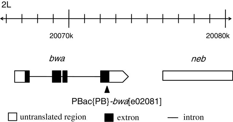
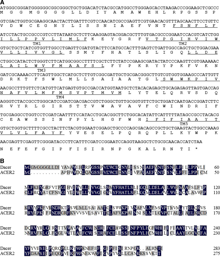
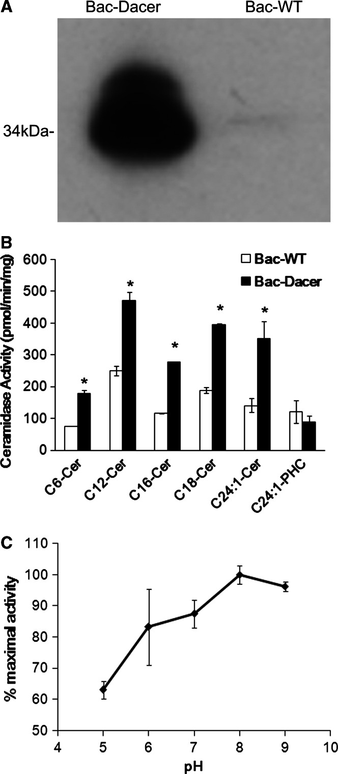
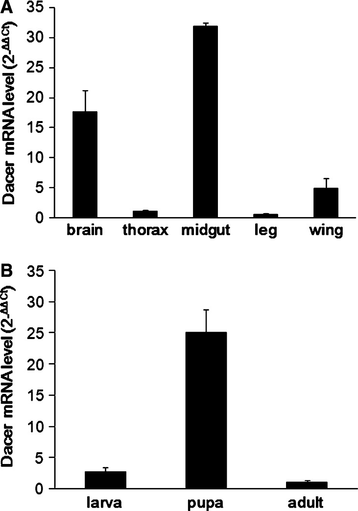
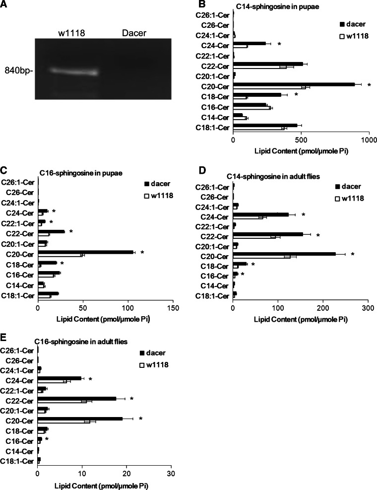
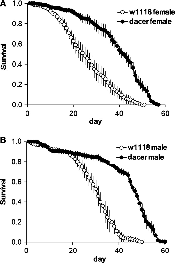
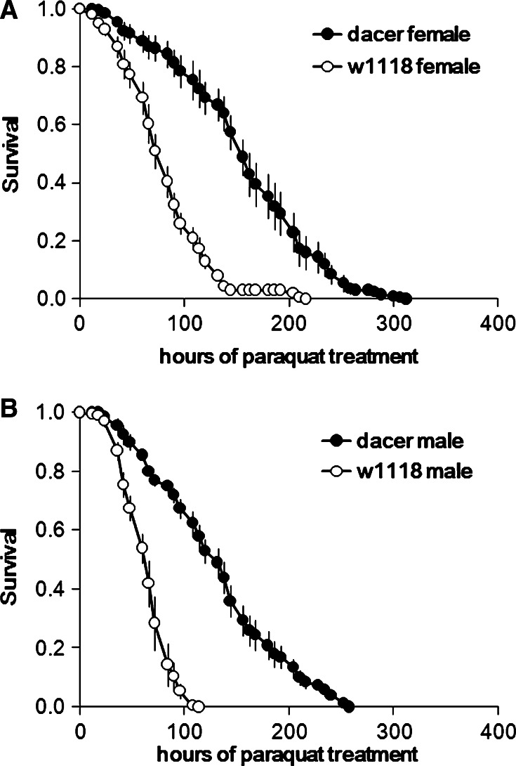
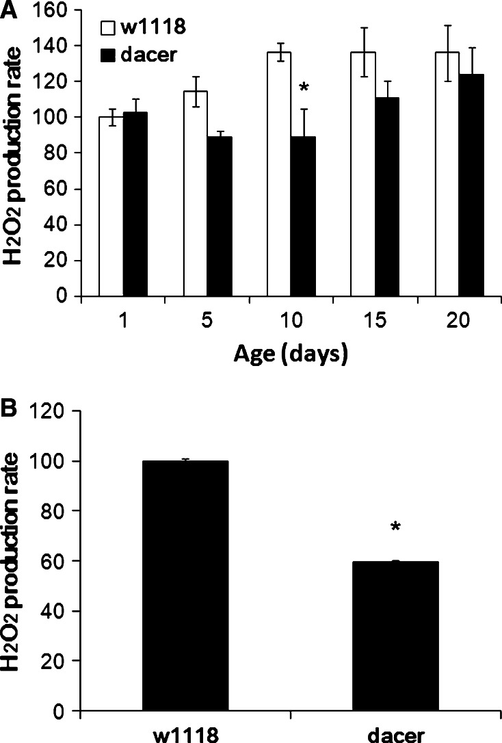
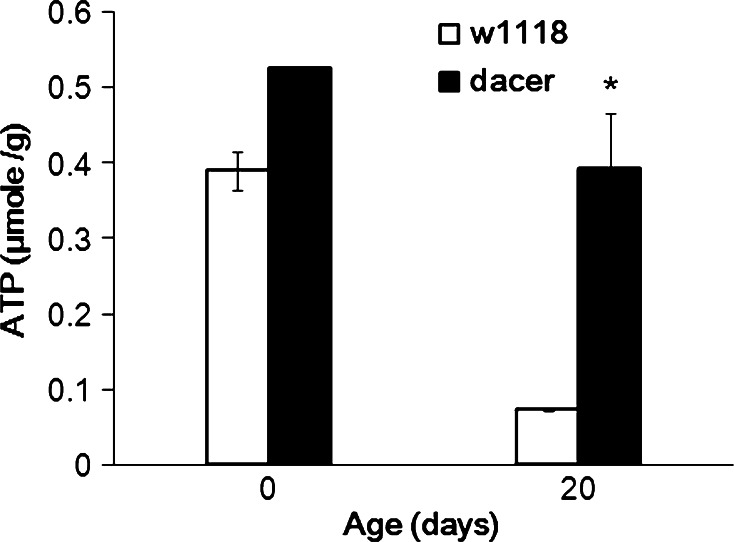
Ceramidases catalyze the hydrolysis of ceramides to generate sphingosine (SPH) and fatty acids, and ceramide metabolism is implicated in various biological responses in Drosophila melanogaster. Here we report the cloning, biochemical characterization, and functional analysis of a Drosophila alkaline ceramidase (Dacer). Dacer, a membrane-bound protein of 284 amino acids, shares homology with yeast and mammalian alkaline ceramidases. Overexpression of Dacer in High Five insect cells increases ceramidase activity in the alkaline pH range, indicating that Dacer is a bona fide alkaline ceramidase. Dacer mRNA is highly expressed in the midgut and at the pupal stage. An inactivation of Dacer by insertional mutagenesis increases the levels of ceramides in both Drosophila pupae and adult flies. Dacer inactivation increases Drosophila pre-adult development time, lifespan, and anti-oxidative stress capacity. Collectively, these results suggest that Dacer plays an important role in the Drosophila development and longevity by controlling the metabolism of ceramides.


Figures

Fig. 1
Fig. 1
Transposon location of the dacer mutant. The Dacer/BWA gene localizes to the 38B2-3 region of the left arm of the second chromosome. A P-element insertion (PBac(43)-bwa e02081) was isolated and was subsequently mobilized to generate a dacer/bwa mutant that carries a transposon within the 4th extron of the Dacer/BWA gene
Fig. 2
Fig. 2
Coding and protein sequences of Dacer. a The Dacer coding sequence (GenBank accession number: AF323976) was cloned from adult Drosophila flies. The amino acid sequence was derived from the coding sequence using the DNAman software. Putative transmembrane domains (TMs1–5) of Dacer were predicted by the pSORT II program. b Protein sequence alignment of Dacer and ACER2 was performed using the DNAman software. Identical amino acid residues among the aligned proteins are shaded in dark grey and similar amino acid residues light grey
Fig. 3
Fig. 3
Dacer encodes alkaline ceramidase activity. a Microsomes were prepared from Bac-Dacer and Bac-WT cells. A portion of the microsomes was subjected to Western blot analysis with the anti-HIS antibody (1:1,000). The relative molecular mass (Mr) (~34 kDa) was estimated according to standard proteins. b Another portion of the above microsomes was assayed for ceramidase activity at pH 9.0 using indicated ceramides as substrates. c Microsomal ceramidase activity was determined using d-e-C16-ceramide as substrate at different pH values. Ceramidase activity of the recombinant Dacer at each pH was computed by subtracting ceramidase activity in Bac-WT microsomes from that in Bac-Dacer microsomes. The Dacer ceramidase activity at pH 8 is highest and set as 100%, and ceramidase activity at other pH values is expressed as % of the maximal activity. All data represent the mean value of three independent experiments performed in duplicate
Fig. 4
Fig. 4
Dacer is highly expressed in the ovary and at the pupal stage. qRT-PCR analysis was performed with cDNAs reversely transcribed from RNA isolated from different Drosophila organs (a) or from flies at different developmental stages (b). Data represent the mean value ± SE of three independent experiments performed in duplicate
Fig. 5
Fig. 5
Dacer inactivation increases Drosophila ceramides. a Total RNA isolated from dacer mutant and control flies was reversely transcribed into cDNAs. The cDNAs were subjected to RT-PCR analysis for Dacer mRNA using the primer pair BWA-5′ and BWA-3′ corresponding to the 5′ and 3′ ends of the coding sequence, respectively. Note that the full-length coding sequence (840 bp) was amplified from controls' RNA, but not from mutants' RNA. b, c. Pupae of mutant and control flies were harvested at 48 h and subjected to ESI/MS/MS analysis for C14SPH- and C16SPH-ceramides. d, e. Adults of mutant and control flies were harvested at day 3 and subjected to ESI/MS/MS analysis for C14SPH- and C16SPH-ceramides. The contents of sphingolipids were normalized to total phospholipids (Pi) in flies. Data represent mean value ± SE of three independent experiments performed in duplicate
Fig. 6
Fig. 6
The dacer mutant flies have enhanced JHAMT activity. Purified JH III acid and [3H]-SAM were dissolved in sodium phosphate solution. CAs were isolated from wandering larvae and homogenized in the sodium phosphate buffer. After centrifugation, the supernatant was transferred to an equal volume of reaction buffer containing JHA and [3H]-SAM. The enzymatic reaction was incubated at 28°C for 90 min and then was stopped by the addition of an equal volume of methanol. The radiolabeled product was extracted with 1 ml of hexane and counted with a liquid scintillation counter. Data represent the mean value ± SE of JHAMT activity (fmol/CA/h). Three independent experiments were performed in duplicate
Fig. 7
Fig. 7
The dacer mutant flies have extended lifespans; 213 mutant and 232 control female as well as 182 mutant and 173 control male eclosion flies were collected. Male and female flies were maintained separately in individual vials on normal food and transferred to fresh vials every 3 days. Survivors were scored every 24 h. Longevity curves of w1118 control flies (open circles) and of dacer mutant flies (filled circles) are shown. For statistical analysis, the mean and maximum lifespan of each strain was calculated from the time (in days) at which survival reached 50 and 10% of the starting population
Fig. 8
Fig. 8
The dacer mutant flies are resistant to oxidative stress. Three-day-old flies were transferred to vials containing standard cornmeal medium added with 20 mM paraquat (20 flies per vial). To measure the survival rate under oxidative stress, the lifespan of control and mutant flies on a paraquat diet was analyzed. Longevity curves of control w1118 flies (open circles) and of dacer mutant flies (filled circles) are shown. For statistical analysis, the mean and maximum lifespan of each strain was calculated from the time (in hours) at which survival reached 50 and 10% of the starting population
Fig. 9
Fig. 9
The dacer mutant flies have decreased ROS production in the mitochondria. a Mutant and control eclosion flies were collected and maintained on normal food. Flies at different ages (1-, 5-, 10-, 15-, and 20-day-old) were kept on ice before isolation of mitochondria. Mitochondria isolated by using Tissue Mitochondria Isolation Kit were incubated at 25°C with 20 mM sn-glycerol 3-phosphate as substrate. Mitochondrial hydrogen peroxide production was measured at 560 nm using the Hydrogen Peroxide Assay Kit. The 100 value refers to control H2O2 production of mitochondria that was obtained from 1-day-old w1118 flies, and other values represent % of the control H2O2 production. b Three-day-old flies were collected and maintained on 20 mM paraquat-containing food. Mitochondria were isolated from dacer and control flies at 36 h after paraquat treatment. Mitochondrial hydrogen peroxide production was measured at 560 nm. The 100 value refers to control H2O2 production of mitochondria that was obtained from w1118 flies, and other values represent % of the control H2O2 production. All data represent the mean value of three independent experiments performed in duplicate
Fig. 10
Fig. 10
The dacer mutant flies have increased ATP levels. ATP levels in the flies were determined by luciferin-luciferase system. Young (day 1) and old flies (day 20) on the normal diet were washed thoroughly in PBS and homogenized. The levels of ATP in homogenates were estimated by luminometer. The ATP levels in control (open bars) and mutant flies (filled bars) are shown. The error bars of the ATP levels in young dacer mutant and old w1118 flies are too small to see

Similar articles

Cited by

References

    1. Acharya U, Acharya JK. Enzymes of sphingolipid metabolism in Drosophila melanogaster . Cell Mol Life Sci. 2005;62:128–142. doi: 10.1007/s00018-004-4254-1. - DOI - PMC - PubMed
    1. Mao CG, Obeid LM. Ceramidases: regulators of cellular responses mediated by ceramide, sphingosine, and sphingosine-1-phosphate. Biochimica et Biophysica Acta (BBA) Mol Cell Biol Lipids. 2008;1781:424–434. doi: 10.1016/j.bbalip.2008.06.002. - DOI - PMC - PubMed
    1. Okino N, Ichinose S, Omori A, Imayama S, Nakamura T, Ito M. Molecular cloning, sequencing, and expression of the gene encoding alkaline ceramidase from Pseudomonas aeruginosa. Cloning of a ceramidase homologue from Mycobacterium tuberculosis. J Biol Chem. 1999;274:36616–36622. doi: 10.1074/jbc.274.51.36616. - DOI - PubMed
    1. Okino N, Tani M, Imayama S, Ito M. Purification and characterization of a novel ceramidase from Pseudomonas aeruginosa . J Biol Chem. 1998;273:14368–14373. doi: 10.1074/jbc.273.23.14368. - DOI - PubMed
    1. Mao CG, Xu RJ, Bielawska A, Obeid LM. Cloning of an alkaline ceramidase from Saccharomyces cerevisiae: an enzyme with reverse (CoA-independent) ceramide synthase activity. J Biol Chem. 2000;275:6876–6884. doi: 10.1074/jbc.275.10.6876. - DOI - PubMed

Publication types

MeSH terms

LinkOut - more resources